
Anesthetic considerations for Ehlers-Danlos syndrome
A perioperative guide for anesthesia providers — and for patients preparing to talk to their anesthesia team.
Every clinical claim on this page is cited to its source below. How we source clinical content.
The short answer
No anesthetic agent is contraindicated in Ehlers-Danlos syndrome, which is not among the conditions that confer malignant hyperthermia susceptibility. The subtype decides the plan: vascular EDS is a different disease from hypermobile EDS. Expect mechanical injury from positioning, adhesives and airway instrumentation, local anesthetic resistance, and bleeding despite normal coagulation studies.
Key takeaways
- The subtype determines the anesthetic. The 2017 classification recognizes 13 types attributed to 19 genes, and additional subtypes have since been proposed.1,31 Hypermobile EDS and vascular EDS require almost entirely different plans.
- Local anesthetic resistance is real and now has controlled evidence. A 2026 randomized cross-over trial found significantly reduced lidocaine effect at 15 and 30 minutes in EDS patients compared with controls.13
- No anesthetic agent is specifically contraindicated. Ehlers-Danlos is not among the conditions associated with malignant hyperthermia susceptibility, which are predominantly disorders of skeletal muscle.26 Volatile agents and succinylcholine are both described as safe in EDS.3
- The predictable injuries are mechanical: joint subluxation from positioning, skin tearing from adhesives, airway mucosal trauma, and hematoma from vascular access.
- Vascular EDS is a different disease. Arterial and hollow-organ rupture drive mortality, median survival is reduced, and elective surgery carries risks the other subtypes do not.18
- Bleeding is common despite normal coagulation studies. A normal PT/INR and platelet count does not rule out a bleeding phenotype.32,33
What Ehlers-Danlos syndrome is, and why the subtype matters more than the label
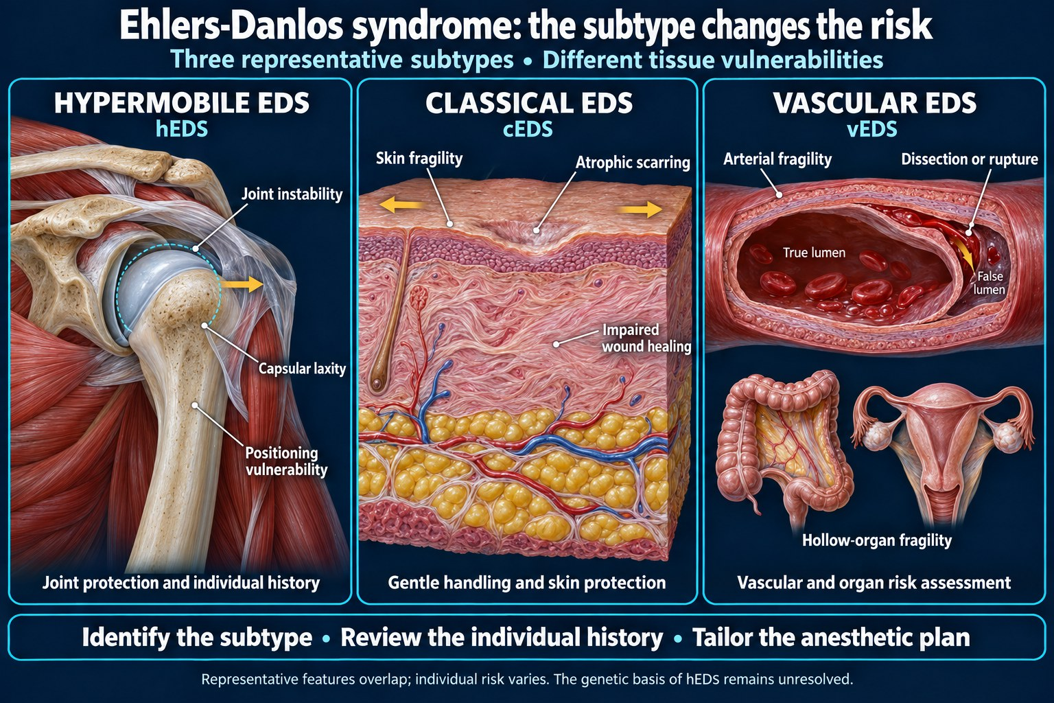
The Ehlers-Danlos syndromes are a group of heritable connective tissue disorders affecting collagen synthesis, processing, or structure. The 2017 international classification recognizes 13 distinct subtypes attributed to variants in 19 genes, each with defined clinical criteria and, for all but one (hypermobile EDS), an identified genetic basis.1 Additional subtypes — including a classical-like type due to AEBP1 variants — have since been described, so the count is best treated as a floor rather than a fixed number.31
This matters clinically because “the patient has EDS” is not enough information to plan an anesthetic. A patient with hypermobile EDS presenting for knee arthroscopy and a patient with vascular EDS presenting for the same operation face almost entirely different risks. The first is a positioning and analgesia problem. The second is a potential arterial catastrophe.
| Subtype | Gene / defect | What changes the anesthetic |
|---|---|---|
| Hypermobile (hEDS) most common | Genetic basis not yet identified | Joint subluxation with positioning, local anesthetic resistance, high rates of POTS, MCAS, gastroparesis and chronic pain. The subtype most anesthesia providers will actually encounter. |
| Classical (cEDS) | COL5A1, COL5A2 | Marked skin fragility and poor wound healing. Adhesive and shear injury risk. Easy bruising. |
| Vascular (vEDS) | COL3A1 | Arterial dissection and rupture, spontaneous bowel and uterine rupture. Every vascular procedure carries risk. Treated as a separate problem below. |
| Kyphoscoliotic (kEDS) | PLOD1, FKBP14 | Restrictive lung disease, scoliosis, and — with PLOD1 — arterial rupture risk. Scleral fragility. |
| Musculocontractural (mcEDS) | CHST14, DSE | Craniofacial features and small mouth complicating intubation, plus documented pneumothorax risk prompting pressure-limited ventilation.2 |
| Classical-like, cardiac-valvular, arthrochalasia, dermatosparaxis, brittle cornea, spondylodysplastic, myopathic, periodontal | Various | Rare. Cardiac-valvular carries severe valve disease; brittle cornea requires eye protection discipline; arthrochalasia carries congenital hip dislocation and severe joint instability; classical-like (tenascin-X deficient) carries GI and tracheal rupture risk.29,30 |
Practical point. Many patients carry a diagnosis of “EDS” made years ago, before genetic testing was available. That diagnosis may be labeled with the numbered types of the older Berlin nosology (type III for hypermobile, type IV for vascular) or with the six named subtypes of the Villefranche nosology.1,3 If the subtype is uncertain, treat the preoperative visit as the place to establish it — and in any patient with a family history of sudden death, arterial rupture, or bowel perforation, assume vascular EDS until proven otherwise.
This is in the app, with the calculators and the drug library — free tier, no card. Get it →
Preoperative assessment
Establish the subtype and the genetics
Ask directly whether genetic testing was performed and what it showed. A confirmed COL3A1 variant changes everything downstream. A clinical diagnosis of hypermobile EDS — which currently has no genetic test — does not carry the same vascular implications.
Obtain previous anesthetic records
This is the highest-yield item in the entire assessment and the most frequently skipped. Prior charts answer questions the physical exam cannot: was the airway difficult, did the block work, was there unexpected bleeding, did the patient have prolonged PACU stays or difficult emergence. Retrospective review of previous anesthesia records has been specifically recommended for this population precisely because the published evidence base is thin enough that individual history outweighs general guidance.3
Cardiac evaluation
Mitral valve prolapse and aortic root dilation occur across several subtypes. Echocardiography is reasonable in any patient with a murmur, a family history of aortic disease, or a subtype with known cardiovascular involvement. Cardiac-valvular EDS carries progressive, severe valve disease and warrants formal cardiology input.
Airway history
Ask about temporomandibular joint problems specifically — TMJ subluxation and dislocation are common, and a patient who dislocates their jaw yawning may dislocate it under laryngoscopy. Ask about neck pain, prior cervical imaging, and any diagnosis of craniocervical instability or Chiari malformation, both of which are over-represented in this population.6
Bleeding history
Standard coagulation testing is frequently normal in patients who nonetheless bleed. Take a structured bleeding history rather than relying on labs — the ISTH bleeding assessment tool has been applied in EDS cohorts, where bleeding scores are markedly higher than in controls32 even though routine coagulation testing is typically normal,33 and point-of-care testing including rotational thromboelastometry and functional platelet assays has been examined against bleeding severity scores in an adult EDS cohort.4
Dysautonomia and POTS
Postural orthostatic tachycardia syndrome co-occurs frequently with hypermobile EDS and materially affects the anesthetic.35 Reported management centers on adequate preoperative hydration, careful positioning during induction, and anticipating hemodynamic lability on emergence.5 Ask about baseline blood pressure, salt and fluid loading regimens, midodrine or fludrocortisone use, and how long the patient has been NPO — prolonged fasting is poorly tolerated.
Mast cell activation syndrome
MCAS is commonly reported alongside hypermobile EDS, though the strength and mechanism of the association remain debated.35 Where it is present, histamine-releasing agents warrant caution, and continuing the patient’s antihistamine regimen (both H1 and H2 blockade) through the perioperative period is reasonable; some clinicians add premedication in patients with a documented reaction history.6
Gastrointestinal function
Gastroparesis and functional GI symptoms are common in hypermobile EDS and are addressed directly in the AGA clinical practice update on GI and autonomic manifestations.35 Consider aspiration risk, and consider gastric ultrasound if the history suggests delayed emptying.
Medication review
Patients with vascular EDS may be taking celiprolol, another beta-blocker, or an angiotensin receptor blocker specifically to reduce arterial event risk.20,27 These should generally be continued. Chronic opioid, NSAID, and anticoagulant use are all common and all relevant.
Airway management
The airway concerns in EDS are mechanical, not anatomical in the conventional difficult-airway sense. Most patients intubate normally. The risk is what the intubation does to them.
Temporomandibular joint
TMJ subluxation is a recognized risk during laryngoscopy and mask ventilation. Minimize mouth opening force, avoid aggressive jaw thrust, and consider that a patient with recurrent TMJ dislocation may benefit from a technique requiring less mouth opening.
Cervical spine
Craniocervical and cervical instability are described in EDS, and cervical instability is a specific concern in vascular EDS.7 New-onset craniocervical instability has been reported in hypermobile EDS after inadvertent neck hyperextension during routine procedures, including intubation.36 Published case management emphasizes avoiding neck hyperextension during intubation and maintaining vigilance for cervical subluxation through emergence and extubation.5 The review most often cited on this point recommends that every EDS patient be considered a candidate for a fiberoptic or video laryngoscopic approach, to minimize manipulation of the head and neck, and says the indication is stronger still where temporomandibular or craniocervical involvement is documented or suspected.6 It does not specify an awake technique, and neither does this page: whether to secure the airway awake is a separate judgment that turns on how much instability has actually been demonstrated.
Mucosal and dental fragility
Airway mucosa tears more readily. Use gentle technique, adequate lubrication, a smaller tube than you might otherwise select, and avoid repeated attempts. In the rare classical-like (tenascin-X deficient) subtype, careful instrumentation is specifically advised because tracheal rupture during intubation and esophageal rupture after probe insertion have been reported.30 Periodontal EDS carries early tooth loss, and dental fragility is worth documenting before instrumentation.
Supraglottic airways
A supraglottic airway has been used successfully in EDS8 and may be preferable where it avoids the mouth opening and cervical movement that laryngoscopy requires, since both are specific vulnerabilities in this population. Balance that against aspiration risk if gastroparesis is present.
Ventilation and pneumothorax
Spontaneous pneumothorax is described in several subtypes. In a reported case of musculocontractural EDS undergoing scoliosis surgery, pressure-controlled ventilation with limited peak inspiratory pressure was used specifically to prevent pneumothorax.2 Lung-protective ventilation with attention to peak pressures is a reasonable default.
Local anesthetic resistance — what the evidence actually shows
This is the single most distinctive anesthetic feature of EDS, the one patients most often report being disbelieved about, and the area where the evidence has changed most recently.
The evidence, in order
1990 — the original observation. A Danish group taking skin biopsies noticed that patients with EDS type III experienced significant pain despite conventional local anesthesia. In the resulting controlled study of 8 patients and 8 controls, both groups achieved analgesia five minutes after intradermal lidocaine, but by 60 minutes the effect had disappeared in the EDS group while persisting in controls. Topical EMLA cream failed to produce adequate analgesia in the EDS patients while controls achieved full anesthesia.9
2005 — the clinical signal. Hakim and colleagues surveyed 172 patients with joint hypermobility syndrome, asking whether a local anesthetic for a dental, surgical or epidural procedure had been as effective as it should have been. A majority — 58% — said it had not, against 21% of controls, an odds ratio of 2.85. The authors call the question simplistic and reliant on recall, and say so themselves. The same letter opens with a smaller, separate Danish series of patients with EDS type III, all of whom reported previous partial or complete failure of local anesthesia in dental or obstetric procedures — and some of whom, the authors note, had been dismissed as hysterical for saying so.10
2019 — the large survey. Among 980 EDS respondents who had received local anesthesia for a dental procedure, 88% recalled inadequate pain prevention, compared with 33% of 249 non-EDS respondents. By agent, the reported success rates were articaine 30%, bupivacaine 25%, and mepivacaine 22% — with lidocaine and procaine reported effective far less often.11 Note what this means: even the best-performing agent worked in fewer than a third of patients.
2022 — resistance across multiple routes. A case report described a laboring patient in whom two technically uneventful epidurals provided no analgesia, the spinal component of a subsequent combined spinal-epidural was also ineffective, and an attempted pudendal block with lidocaine had no effect. General anesthesia was ultimately required for cesarean delivery. The authors described it as the first report of local anesthetic resistance by three distinct routes in one patient.12
2026 — the randomized trial. The strongest evidence to date. In a randomized cross-over design, 135 participants (91 with EDS, 44 healthy controls) received subcutaneous saline and lidocaine at separate forearm sites, with sensation assessed by standardized monofilament. At five minutes there was no difference between groups. At 15 and 30 minutes, adequate anesthesia was present in 60% versus 84% (p = 0.006) and 53% versus 80% (p = 0.003) respectively.13
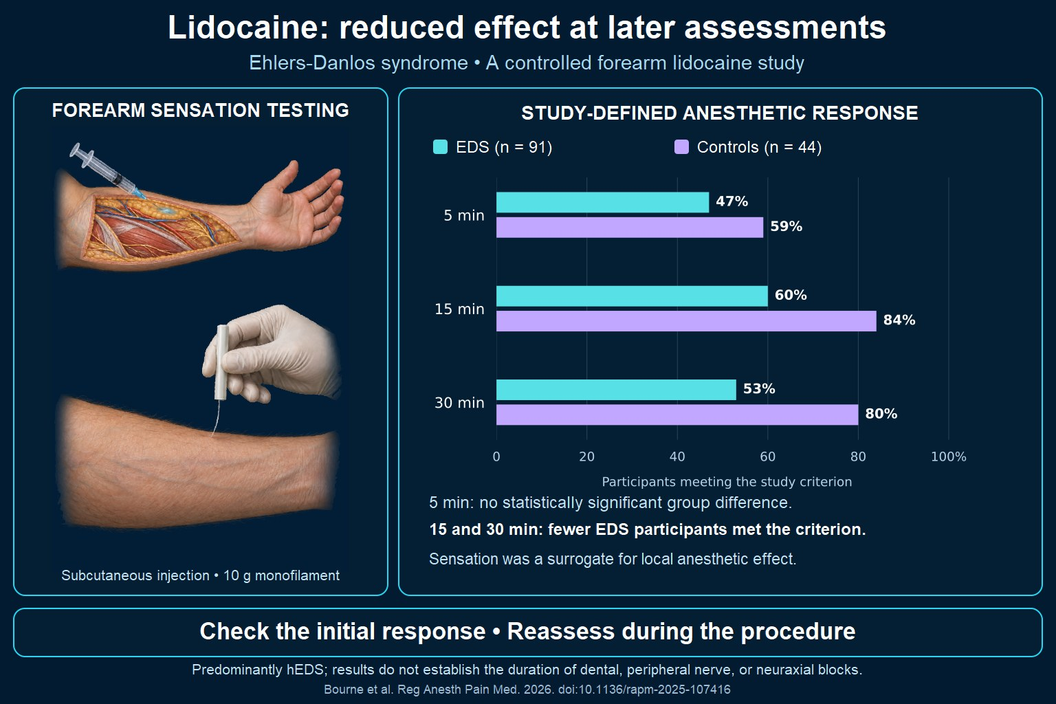
The clinical shape of the finding: onset is normal, duration is short. Patients are not failing to become numb — they are becoming numb and then waking up mid-procedure. That distinction matters, because it means a block that appears to work at the five-minute check may not hold, and it explains why patient reports have historically been dismissed by clinicians who tested the block and found it adequate.
What to do about it
- Believe the history. A patient who tells you lidocaine does not work for them is describing a phenomenon with controlled evidence behind it.13
- Ask which agents have worked before. Response appears agent-specific, and the survey data favor articaine, bupivacaine, and mepivacaine over lidocaine and procaine.11
- Prefer longer-acting agents where the procedure allows, since the failure mode is duration rather than onset.
- Re-test the block before incision, and again later — not only at five minutes.
- Have a plan B stated out loud before starting, especially for awake procedures and labor analgesia. The patient should know what happens if the block fails.
- Do not exceed maximum safe doses chasing an inadequate block. Resistance is not a reason to escalate past toxic thresholds; it is a reason to change technique. Ceilings for every agent are tabulated at local anesthetic maximum doses.
Important distinction. Local anesthetic resistance is not local anesthetic allergy. True amide allergy is rare, and a history of a “reaction” at the dentist is far more often epinephrine effect, vasovagal syncope, or preservative sensitivity. Both have been reported in EDS, and they require completely different management — one calls for a different agent or technique, the other for allergy referral and testing.14
Regional and neuraxial anesthesia
Neuraxial technique in EDS is a subtype decision rather than a single answer, and the cited recommendation differs depending on which subtype you are dealing with.
In vascular EDS, the recommendation is to avoid it. Wiesmann and colleagues recommend avoiding neuraxial blockade in vascular EDS, reasoning that there is no clear benefit sufficient to justify the potential risk when compared with a planned general anesthetic. They extend the same recommendation to peripheral nerve blocks in this subtype on bleeding grounds. They acknowledge that successful spinal and epidural anesthetics in vascular EDS have been reported, but caution that underreporting bias makes those reports a poor basis for generalizing, and state explicitly that they should not be read as a carte blanche. The Orphanet emergency guidance for vascular EDS and older obstetric guidance are cited as taking the same restrictive position.3
In other EDS subtypes, neuraxial blockade is described as feasible, with the qualification that the benefits and risks be worked through explicitly with the patient and the team. The factors named for that discussion are the specific subtype, bleeding diathesis, spinal pathology, and any history of hematoma or spontaneous organ rupture, weighed against the real benefits of a neuraxial technique: early mobilization, pain control, and being awake for delivery.3 Successful use, including in obstetrics, is documented.3,15
Bleeding and hematoma
Increased epidural hematoma risk has been raised specifically in the context of vascular-type EDS, alongside the general concern for excessive bleeding.7 This is the principal reason the recommendation against neuraxial technique in that subtype exists. Vascular complications, chiefly hematoma, are also documented in several nominally “nonvascular” subtypes, so a bleeding phenotype deserves weight wherever it is present.34 In hypermobile EDS without a bleeding phenotype the concern is smaller but not zero, and large trials estimating the risk of perforation, nerve injury, and hematoma formation in patients with tissue fragility have not been done.3
Technical difficulty and dural puncture
Connective tissue laxity may make landmarks less reliable and the epidural space harder to identify. Scoliosis, previous spinal surgery, and other spine pathology should be ruled out before a neuraxial technique is planned, and pre-procedural ultrasound or MRI has been suggested for that purpose.3 Ultrasound guidance in practice is reasonable and has been described: a recent case report of cesarean delivery in classical EDS used ultrasound-guided spinal anesthesia as part of a coordinated multidisciplinary plan.15
Tarlov cysts are a feature of classic, hypermobile, and kyphoscoliotic EDS. Most sit between S1 and S4 and represent only a relative contraindication to spinal or lumbar and thoracic epidural technique, though thoracic cysts have been observed in hypermobile EDS.3
The sources diverge on post-dural puncture headache. One states that risk is increased in this population.6 The other argues that a higher rate might be expected on tissue-fragility grounds but that no relevant studies exist, and notes that the published neuraxial literature in EDS, mostly obstetric, contains a single reported PDPH.3 Heritable connective tissue disorders, EDS among them, are also over-represented in patients with spontaneous spinal CSF leaks and orthostatic headache,38 which adds to the plausibility of a higher PDPH rate without establishing one. Treat elevated risk as plausible and worth consenting for rather than established. If PDPH does occur, an epidural blood patch remains available.3
Peripheral nerve blocks
Peripheral nerve blocks are not recommended in vascular EDS, on the same bleeding grounds as neuraxial technique. In other subtypes they are advised on a case-by-case, shared-decision basis.3 Where they are appropriate they are often desirable: the frequency of difficult airway status across EDS subtypes is itself an argument for regional technique in limb surgery, and the opioid-sparing benefit matters in a population with high chronic pain prevalence. Tissue scarring may theoretically impede local anesthetic spread, and ultrasound guidance is recommended both for that reason and to reduce vascular puncture.3
Apply the local anesthetic resistance caveats throughout: verify the block, expect shorter duration than usual, and consider a catheter rather than a single shot where the procedure warrants it.
General anesthesia
The most common misconception is that EDS carries malignant hyperthermia risk. It is not among the conditions associated with malignant hyperthermia susceptibility, which are predominantly disorders of skeletal muscle.26 The anesthesia literature on EDS reads accordingly: there is no evidence that the diagnosis itself alters anesthetic pharmacokinetics or pharmacodynamics, balanced anesthesia with volatile agents, nitrous oxide, or TIVA are all described as acceptable, and both depolarizing and non-depolarizing neuromuscular blocking agents are described as safe.3
Agent selection
There are no subtype-specific agent contraindications. Where MCAS is present, caution with histamine-releasing agents is advised, and induction agents producing minimal hemodynamic change, such as etomidate or ketamine, may be preferred.6
Neuromuscular blockade
Succinylcholine is not contraindicated by EDS itself. The standard exception applies: avoid depolarizing agents in patients who are immobilized, where upregulation of extrajunctional receptors creates hyperkalemia risk independent of the connective tissue diagnosis. Because some subtypes involve muscle weakness — myopathic EDS most obviously — quantitative neuromuscular monitoring before emergence is specifically advised.3
Opioid response
Unpredictable opioid response is widely reported in this population, though the evidence is largely anecdotal.6 Many patients have substantial chronic opioid exposure and correspondingly elevated requirements. Multimodal, opioid-sparing strategies have been described specifically for hypermobile EDS, including in patients with craniocervical instability and severe chronic pain undergoing major surgery (occipito-cervical fixation).37
Positioning, skin, and the injuries that actually happen
If a patient with EDS is harmed perioperatively, this is the most likely mechanism. The injuries are preventable and they are mechanical.
Joint subluxation and dislocation
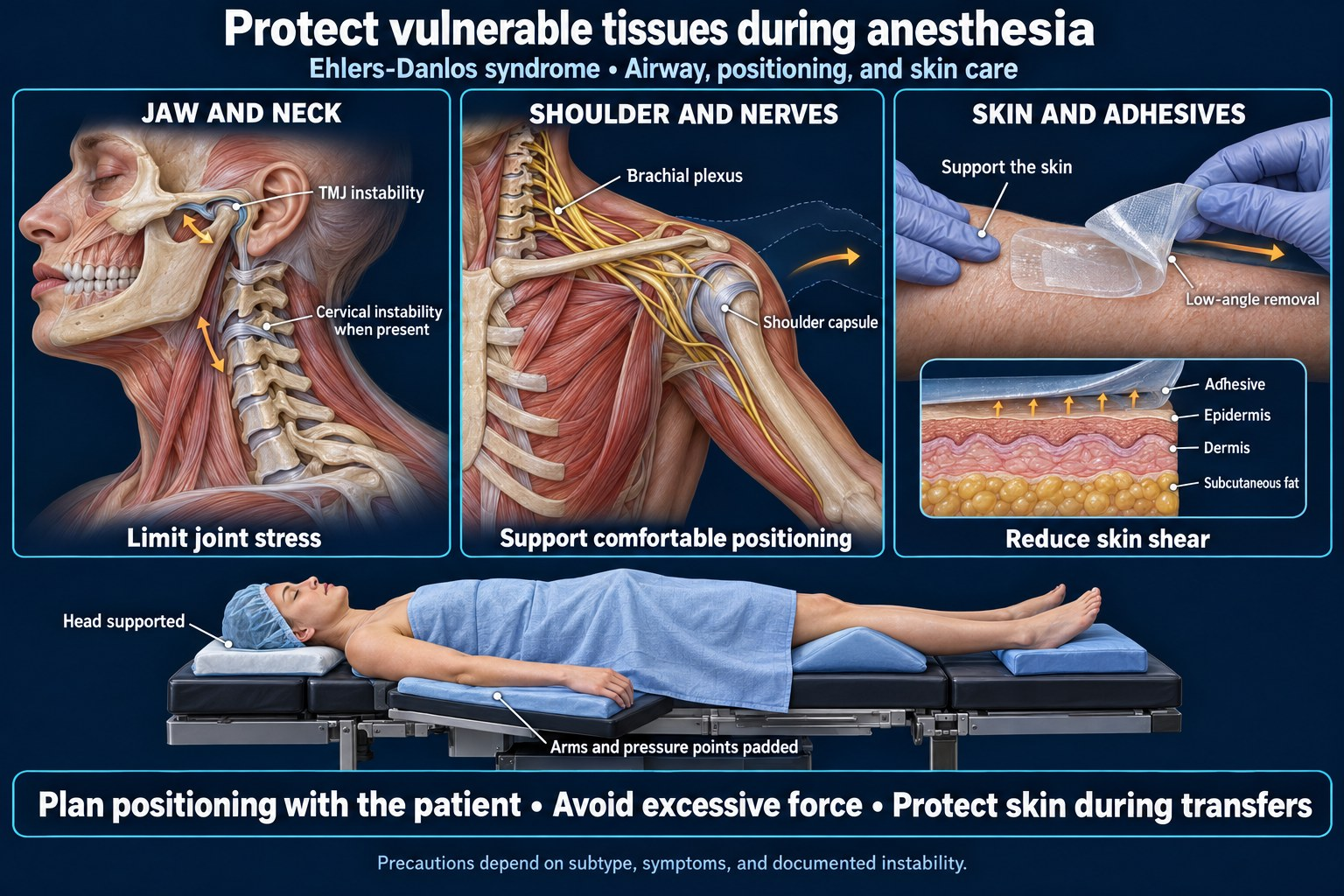
Joints that dislocate during ordinary activity will dislocate under anesthesia, when protective muscle tone is gone and the patient cannot report discomfort. Position the patient before induction where possible and ask them to confirm the position is comfortable and sustainable. Avoid extremes of abduction, rotation, and traction. Move the patient as a unit, with enough hands.3
Peripheral nerve injury
A case of perioperative brachial plexus injury caused by upper extremity hyperabduction in the prone position has been reported specifically in a patient with EDS.16 Standard positioning limits should be treated as maximums rather than targets.
Skin and adhesive injury
Skin tears from shear force and from medical adhesives are a genuine risk, particularly in classical and dermatosparaxis subtypes. Reported mitigation includes low-rebound mattresses to reduce shear and external pressure, and barrier films to prevent medical adhesive–related skin injury.2 Wound healing is delayed: deep or severe wounds in any EDS type should be closed with tension-free sutures placed generously in layers, left in for about twice as long as usual or longer, with tape over the repair to limit stretching.39 Practically: use paper tape or silicone-based dressings, avoid aggressive adhesive removal, consider a barrier film under any adhesive, and pad generously.
Tourniquets
Tourniquet use warrants explicit discussion with the surgeon in subtypes with vascular fragility, given the risk of hematoma, compartment syndrome, and diffuse bleeding.3
Eyes
Brittle cornea syndrome and scleral fragility in kyphoscoliotic EDS make eye protection non-optional. Avoid any pressure on the globe.1
Bleeding and hemostasis
Easy bruising and prolonged bleeding are common across subtypes and frequently occur with entirely normal routine coagulation studies. The defect is largely in the vessel wall and perivascular connective tissue rather than in the coagulation cascade, though functional platelet abnormalities are also reported.33
- Do not be reassured by a normal PT/INR and platelet count. Take the history; ISTH-BAT bleeding scores are markedly elevated in EDS cohorts despite normal routine coagulation studies.32,33
- Desmopressin has been studied for EDS-associated bleeding symptoms and is a reasonable consideration in patients with a documented phenotype.17
- Tranexamic acid is a reasonable adjunct in procedures with meaningful expected blood loss.
- Viscoelastic testing (TEG or ROTEM) has been used to guide hemostatic management, including in a reported case of severe perioperative bleeding during scoliosis surgery in musculocontractural EDS.2
- Vascular access is itself a bleeding risk. Expect hematoma from IV attempts, use ultrasound, and minimize attempts.
Vascular EDS: a different problem entirely
This section is not interchangeable with the rest of the page. Vascular EDS carries a risk of sudden arterial and hollow-organ rupture that the other subtypes do not, and management decisions that are routine elsewhere may be dangerous here.
Vascular EDS results from pathogenic variants in COL3A1, which encodes type III procollagen — a major structural protein of vessel walls and hollow organs. Estimated frequency is between 1 in 50,000 and 1 in 200,000.18 Characteristic features include thin translucent skin, easy bruising, distinctive facial features, arterial dissection and rupture, and spontaneous rupture of bowel or the gravid uterus.18 Roughly a quarter of patients experience a vascular complication before age 20, rising to about 80% by age 40,19 and median survival is about 51 years.18
Anesthetic implications
- Vascular access is both difficult and hazardous. Arterial puncture carries real risk. Where invasive monitoring is genuinely required, weigh it explicitly rather than placing lines by default, and use ultrasound.3,18
- Spontaneous arterial rupture can occur perioperatively — a documented concern independent of surgical manipulation.7
- Blood pressure control is the central medical intervention.22,41 Avoid hypertensive surges at laryngoscopy, emergence, and extubation. Treat coughing and straining as hemodynamic events.
- Cervical instability and airway trauma risk are specifically described in this subtype.7
- Bowel and uterine rupture may be the presenting surgical problem, which means these patients can arrive as emergencies without a known diagnosis.18
Celiprolol and medical therapy
The BBEST trial — a prospective randomized open, blinded-endpoint study — reported a substantially reduced hazard of arterial events with celiprolol, a beta-1 antagonist with beta-2 agonist vasodilatory activity, titrated up to 400 mg daily.20 The result has been questioned because a proportion of participants lacked genetically confirmed COL3A1 variants, and subsequent cohort studies have reported both tolerability limits and continued fatal events on treatment.21 The 2022 ACC/AHA aortic disease guideline stated that there were no studies showing a benefit of angiotensin receptor blockers in vascular EDS,28 but a 2025 randomized, double-blind, placebo-controlled trial subsequently found that adding irbesartan to background celiprolol reduced new arterial events over two years.27,42 Current practice emphasizes strict blood pressure control with beta-blockers and angiotensin receptor blockers, alongside surveillance imaging and lifestyle modification.19,22
Availability note. Celiprolol is not marketed in the United States, and available data were considered insufficient for FDA approval. US clinicians commonly substitute another beta-blocker — sometimes one with vasodilatory properties — or rely on an ARB, so a US vascular EDS patient may be on a different regimen while pursuing the same goal of strict blood pressure control.28 Celiprolol is not available in Canada either.40
For the anesthesia provider the practical point is narrower: these medications should generally be continued perioperatively, and their presence in the medication list is itself a signal that the patient has confirmed vascular EDS.
Surgery in vascular EDS
Historically, surgical intervention was avoided until complications developed, on the reasoning that tissue fragility made operating hazardous. That position has been modified in selected cases, and endovascular approaches previously avoided in connective tissue disorders are increasingly reported as feasible with careful selection and meticulous technique.23,24 This is a decision for a multidisciplinary team at a center with experience, with shared decision-making, not a decision to be made intraoperatively.22,28,42
Obstetric considerations
Pregnancy in EDS carries elevated risk of prematurity, hemorrhage, and maternal morbidity. In vascular EDS specifically, uterine rupture and arterial rupture during pregnancy and delivery are recognized causes of maternal death, and early planned cesarean delivery has been advocated to avoid severe lacerations.25
A case report of elective cesarean delivery in classical EDS describes the shape of good practice: a multidisciplinary plan built in advance with cardiology, hematology, endocrinology and pulmonology input, ultrasound-guided spinal anesthesia, and extended post-anesthesia observation.15 The recurring theme across the obstetric literature is that the plan should exist weeks before labor, not on arrival.
Labor analgesia deserves particular attention given the local anesthetic resistance data — the multi-route failure case described above occurred in a laboring patient, and the fallback was general anesthesia for cesarean delivery.12
Postoperative care
- Pain control is frequently inadequate in this population, reflecting both local anesthetic resistance and chronic pain with opioid tolerance. Plan multimodal analgesia deliberately rather than reactively.
- Watch for occult bleeding. A high index of suspicion is warranted given the combination of vessel fragility and normal-appearing coagulation studies.6
- Hemodynamic instability may persist where POTS or dysautonomia is present. Extended monitored observation is reasonable.5,15
- Wound healing is delayed. Sutures are typically left in place about twice as long as usual or longer, and layered, tension-free closure is recommended.39
- Post-dural puncture headache risk may be elevated after neuraxial technique; the evidence is limited.3,6
- Joint protection continues into recovery — transfers from the operating table to the bed and from bed to chair are dislocation opportunities.
A practical preoperative checklist
| Item | Why |
|---|---|
| Which subtype, and was it genetically confirmed? | Determines whether this is a positioning problem or a vascular one |
| Previous anesthetic records obtained | Highest-yield single item; the evidence base is thin enough that personal history outweighs it |
| Which local anesthetics have worked before? | Response is agent-specific and resistance is common |
| Structured bleeding history taken | Routine coagulation studies are often normal in patients who bleed |
| Echocardiogram if indicated | MVP, aortic root dilation, valve disease by subtype |
| TMJ and cervical spine history | Both are injured by routine airway management |
| POTS status, hydration plan, NPO duration minimized | Prolonged fasting is poorly tolerated |
| MCAS status and antihistamine regimen | Determines agent selection |
| Gastroparesis assessment | Aspiration risk |
| Positioning rehearsed awake | Protective tone is absent under anesthesia |
| Adhesive plan — barrier film, silicone or paper tape | Skin tearing is a real and preventable injury |
| Plan B stated aloud for any awake or regional technique | Block failure should not be improvised around |
Frequently asked questions
Is anesthesia safe for someone with Ehlers-Danlos syndrome?
Yes, with planning. Safe anesthesia can be provided with adequate preparation and appropriate resources.3 The risks are largely predictable and preventable: joint injury from positioning, skin damage from adhesives, airway trauma, bleeding, and inadequate local anesthesia. Vascular EDS carries additional and more serious risks that require specialist management.
Does local anesthetic really not work in EDS, or is that a myth?
It is real and it now has controlled evidence. A 2026 randomized cross-over trial found significantly reduced lidocaine effectiveness at 15 and 30 minutes in EDS patients compared with healthy controls, though not at 5 minutes.13 Earlier work showed the same pattern — normal onset, shortened duration.9 Survey data found 88% of EDS respondents reported inadequate pain prevention during dental procedures versus 33% of non-EDS respondents.11
Which local anesthetic works best in Ehlers-Danlos syndrome?
Survey data suggest articaine performs best, followed by bupivacaine and mepivacaine, with lidocaine and procaine reported effective far less often. However, even the best-performing agent was reported effective in only about 30% of EDS respondents.11 The most useful guide is the individual patient’s own history of what has worked.
Can people with EDS have an epidural or spinal?
Usually yes, but it depends on the subtype. In most types of EDS, neuraxial anesthesia is not contraindicated and has been used successfully, including a reported case of elective cesarean delivery under ultrasound-guided spinal anesthesia in classical EDS.3,15 Vascular EDS is the exception: the published recommendation is to avoid spinal and epidural anesthesia in that subtype, because the bleeding and hematoma risk is not offset by a clear enough benefit compared with a planned general anesthetic. Successful cases have been reported in vascular EDS, but those authors caution that such reports are subject to underreporting bias and should not be treated as general permission.3
Where a neuraxial technique is appropriate, the considerations are bleeding and hematoma risk, potentially difficult landmarks and any underlying spine pathology, a possibly increased risk of post-dural puncture headache, and the real possibility that the block will be less effective or shorter-lived than expected.3,6,12 The decision is worth making in advance with your anesthesia team rather than on the day, and if you have vascular EDS or don’t know your subtype, say so early.
Is Ehlers-Danlos syndrome linked to malignant hyperthermia?
No. Ehlers-Danlos is not among the conditions associated with malignant hyperthermia susceptibility, which are predominantly disorders of skeletal muscle — central core disease, King-Denborough syndrome, multiminicore disease, the muscular dystrophies and the myotonias among them.26 The practical consequence shows up in the anesthesia guidance for EDS itself: the triggering drugs, volatile agents and succinylcholine, are both described as safe, and there is no evidence that the diagnosis alters anesthetic pharmacokinetics or pharmacodynamics.3 This is one of the most common misconceptions about the condition.
What should I tell my anesthesia provider before surgery if I have EDS?
Your subtype and whether it was genetically confirmed; which local anesthetics have and have not worked for you; any history of joint dislocation and which joints; whether your skin tears with tape; whether you have POTS, MCAS, or gastroparesis; your bleeding history including dental extractions and childbirth; any neck or jaw problems; and — most usefully — records from any previous anesthetic.
Is surgery riskier with vascular EDS?
Yes, substantially. Vascular EDS carries risk of arterial dissection and rupture and spontaneous bowel or uterine rupture, with roughly 25% of patients experiencing a vascular complication before age 20 and about 80% by age 40,19 and reduced median survival.18 Elective surgery decisions should involve a multidisciplinary team with experience in the condition.
References
- Malfait F, Francomano C, Byers P, et al. The 2017 international classification of the Ehlers-Danlos syndromes. Am J Med Genet C Semin Med Genet. 2017;175(1):8–26. doi:10.1002/ajmg.c.31552
- Wakabayashi R, Tanaka S, Tsuchiyama K, et al. Anesthetic management of a patient with musculocontractural Ehlers-Danlos syndrome undergoing scoliosis surgery. JA Clin Rep. 2020;6:41. doi:10.1186/s40981-020-00352-5
- Wiesmann T, Castori M, Malfait F, Wulf H. Recommendations for anesthesia and perioperative management in patients with Ehlers-Danlos syndrome(s). Orphanet J Rare Dis. 2014;9:109. doi:10.1186/s13023-014-0109-5
- Lam AL, Mittal N, Vinod M, et al. The association of point-of-care coagulation testing with bleeding symptoms in patients with Ehlers-Danlos syndrome: an exploratory cross-sectional study. Can J Anesth. 2025. doi:10.1007/s12630-025-03048-5. PMID 41419706. Toronto General Hospital GoodHope EDS Clinic cohort; ROTEM, PlateletWorks and ISTH-BAT.
- Laserna A, Nishtar M, Vidovich C, Borovcanin Z. Perioperative management of Ehlers-Danlos type III syndrome associated with postural orthostatic tachycardia in patients undergoing general anesthesia. Cureus. 2021;13(11):e19311. doi:10.7759/cureus.19311
- Chopra P, Bluestein L. Perioperative care in patients with Ehlers Danlos syndromes. Open Journal of Anesthesiology. 2020;10(1):13–29. doi:10.4236/ojanes.2020.101002. Review of coexisting conditions including dysautonomia, MCAS, Chiari malformation, craniocervical instability, gastroparesis, and altered local anesthetic response.
- Lane D. Anaesthetic implications of vascular type Ehlers-Danlos syndrome. Anaesth Intensive Care. 2006;34(4). doi:10.1177/0310057X0603400412. PMID 16913351
- Daneshvar H, Abtahi D. Perioperative anesthesia management in a patient with Ehlers-Danlos syndrome: a case report of scleral buckling surgery. Arch Anesth Crit Care. 2026. doi:10.18502/aacc.v12i3.21319.
- Arendt-Nielsen L, Kaalund S, Bjerring P, Høgsaa B. Insufficient effect of local analgesics in Ehlers Danlos type III patients (connective tissue disorder). Acta Anaesthesiol Scand. 1990;34(5):358–361. doi:10.1111/j.1399-6576.1990.tb03103.x. PMID 2389651.
- Hakim AJ, Grahame R, Norris P, Hopper C. Local anaesthetic failure in joint hypermobility syndrome. J R Soc Med. 2005;98(2):84–85. doi:10.1177/014107680509800222. PMID 15684369. PMC1079398.
- Schubart JR, Schaefer E, Janicki P, et al. Resistance to local anesthesia in people with the Ehlers-Danlos syndromes presenting for dental surgery. J Dent Anesth Pain Med. 2019;19(5):261–270. PMID 31723666
- Jacobs A, et al. Resistance to local anaesthetics administered via epidural, intrathecal and pudendal injections. Anaesth Rep. 2022;10(2):e12205. doi:10.1002/anr3.12205
- Bourne KM, Thai S, Lei LY, et al. Patients with Ehlers-Danlos syndrome experience reduced effectiveness of lidocaine local anesthetic: a randomized cross-over clinical trial. Reg Anesth Pain Med. 2026. doi:10.1136/rapm-2025-107416. PMID 41775498. Note: this paper reports 44% versus 91% at 30 minutes in its abstract and 53% versus 80% in its results text, both at p = 0.003. The results-text figures are the ones quoted above.
- Cesare AE, Rafer LC, Myler CS, Brennan KB. Anesthetic management for Ehlers-Danlos syndrome, hypermobility type complicated by local anesthetic allergy: a case report. Am J Case Rep. 2019;20:39–42. doi:10.12659/AJCR.912799
- Aljuba YM, Shatalin D, Ronenson A, Grenader A, Ioscovich A. Anesthetic management of a pregnant patient with Ehlers-Danlos syndrome undergoing elective cesarean delivery: a case report. Cureus. 2025;17(8):e89400. doi:10.7759/cureus.89400
- Ohashi N, Furutani K, Ishii H, Baba H. Perioperative brachial plexus injury caused by hyperabduction of the upper extremity in a patient with Ehlers-Danlos syndrome in the prone position. Masui. 2012;61(6):626–628. PMID 22746029.
- Mast KJ, Nunes ME, Ruymann FB, Kerlin BA. Desmopressin responsiveness in children with Ehlers-Danlos syndrome associated bleeding symptoms. Br J Haematol. 2009;144(2):230–233. doi:10.1111/j.1365-2141.2008.07446.x. PMID 19036109.
- Byers PH, Belmont J, Black J, et al. Diagnosis, natural history, and management in vascular Ehlers-Danlos syndrome. Am J Med Genet C Semin Med Genet. 2017;175(1):40–47. doi:10.1002/ajmg.c.31553
- Jaiswal M, Mansoorshahi S, Zachariah S, et al. Varied presentations of arterial events in vascular Ehlers-Danlos syndrome. JACC Case Rep. 2025;30(32). doi:10.1016/j.jaccas.2025.105424. PMID 40938243
- Ong KT, Perdu J, De Backer J, et al. Effect of celiprolol on prevention of cardiovascular events in vascular Ehlers-Danlos syndrome: a prospective randomised, open, blinded-endpoints trial (BBEST). Lancet. 2010;376(9751):1476–1484. doi:10.1016/S0140-6736(10)60960-9. PMID 20825986.
- Baderkhan H, Wanhainen A, Stenborg A, et al. Celiprolol treatment in patients with vascular Ehlers-Danlos syndrome. Eur J Vasc Endovasc Surg. 2021;61(2):326–331. doi:10.1016/j.ejvs.2020.10.020. PMID 33223285.
- Lian T, Bhandari A, Shalhub S. What every vascular surgeon should know about vascular Ehlers-Danlos syndrome. Ann Vasc Surg. 2026;129:302–306. doi:10.1016/j.avsg.2026.03.032. PMID 41905459.
- Khoury MK, Eagleton MJ. Iliac artery aneurysm endoleak management in a patient with vascular Ehlers-Danlos syndrome. J Vasc Surg Cases Innov Tech. 2024. doi:10.1016/j.jvscit.2023.101401
- Chu LC, Johnson PT, Dietz HC, et al. Vascular complications of Ehlers-Danlos syndrome: CT findings. AJR Am J Roentgenol. 2012;198(2):482–487. doi:10.2214/AJR.11.6603. PMID 22268198
- Bowen JM, Hernandez M, Johnson DS, et al. Diagnosis and management of vascular Ehlers-Danlos syndrome: experience of the UK national diagnostic service, Sheffield. Eur J Hum Genet. 2023;31:749–760. doi:10.1038/s41431-023-01343-7
- Associated conditions. Malignant Hyperthermia Association of the United States. mhaus.org/healthcare-professionals/miscellaneous/associated-conditions. Accessed September 2026. Lists the conditions associated with malignant hyperthermia susceptibility — central core disease, King-Denborough syndrome, multiminicore disease, the muscular dystrophies, the myotonias, the periodic paralyses, osteogenesis imperfecta and carnitine palmityl transferase deficiency. Ehlers-Danlos syndrome is not among them.
- Jeunemaitre X, Mousseaux E, Frank M, et al. Efficacy of irbesartan in celiprolol-treated patients with vascular Ehlers-Danlos syndrome. Circulation. 2025;151(10):686–695. doi:10.1161/CIRCULATIONAHA.124.072849. PMID 39906986.
- Isselbacher EM, Preventza O, Hamilton Black J III, et al. 2022 ACC/AHA guideline for the diagnosis and management of aortic disease. J Am Coll Cardiol. 2022;80(24):e223–e393. doi:10.1016/j.jacc.2022.08.004
- Green C, Ghali N, Akilapa R, et al. Classical-like Ehlers-Danlos syndrome: a clinical description of 20 newly identified individuals with evidence of tissue fragility. Genet Med. 2020;22(10):1576–1582. doi:10.1038/s41436-020-0850-1
- van Gurp JE, Lechner RL, Micha D, et al. Tenascin-X deficiency causing classical-like Ehlers-Danlos syndrome type 1 in humans is a significant risk factor of gastrointestinal and tracheal ruptures. Clin Transl Gastroenterol. 2025;16(3):e00821. doi:10.14309/ctg.0000000000000821. PMID 39807789.
- Blackburn PR, Xu Z, Tumelty KE, et al. Bi-allelic alterations in AEBP1 lead to defective collagen assembly and connective tissue structure resulting in a variant of Ehlers-Danlos syndrome. Am J Hum Genet. 2018;102(4):696–705. doi:10.1016/j.ajhg.2018.02.018. PMID 29606302.
- Kumskova M, Flora GD, Staber J, Lentz SR, Chauhan AK. Characterization of bleeding symptoms in Ehlers-Danlos syndrome. J Thromb Haemost. 2023;21(7):1824–1830. doi:10.1016/j.jtha.2023.04.004. PMID 37179130.
- Artoni A, Bassotti A, Abbattista M, et al. Hemostatic abnormalities in patients with Ehlers-Danlos syndrome. J Thromb Haemost. 2018;16:2425–2431. doi:10.1111/jth.14310
- D’hondt S, Van Damme T, Malfait F. Vascular phenotypes in nonvascular subtypes of the Ehlers-Danlos syndrome: a systematic review. Genet Med. 2018;20:562–573. doi:10.1038/gim.2017.138. PMID 28981071.
- Aziz Q, Harris LA, Goodman BP, Simrén M, Shin A. AGA clinical practice update on GI manifestations and autonomic or immune dysfunction in hypermobile Ehlers-Danlos syndrome: expert review. Clin Gastroenterol Hepatol. 2025;23(8):1291–1302. doi:10.1016/j.cgh.2025.02.015
- Lee C, Sutherland S, Chopra P. Craniocervical instability after inadvertent neck hyperextension in Ehlers-Danlos syndrome: a retrospective case series and literature review. BMC Neurol. 2026;26:291. doi:10.1186/s12883-026-04801-z. PMID 41872801.
- Ramírez-Paesano C, Rodiera Clarens C, Sharp Segovia A, et al. Perioperative opioid-minimization approach as a useful protocol in the management of patients with Ehlers-Danlos syndrome-hypermobility type, craniocervical instability and severe chronic pain who are to undergo occipito-cervical fixation. Orphanet J Rare Dis. 2023;18:214. doi:10.1186/s13023-023-02829-9
- Schievink WI, Gordon OK, Tourje J. Connective tissue disorders with spontaneous spinal cerebrospinal fluid leaks and intracranial hypotension: a prospective study. Neurosurgery. 2004;54(1):65–70. doi:10.1227/01.neu.0000097200.18478.7b. PMID 14683542.
- Malfait F, Castori M, Francomano CA, et al. The Ehlers–Danlos syndromes. Nat Rev Dis Primers. 2020;6(1):64. doi:10.1038/s41572-020-0194-9. Tension-free layered closure, sutures left about twice as long, tape over the repair.
- Wong GW, Boyda HN, Wright JM. Blood pressure lowering efficacy of partial agonist beta blocker monotherapy for primary hypertension. Cochrane Database Syst Rev. 2014;(11):CD007450. doi:10.1002/14651858.CD007450.pub2. Notes that celiprolol is not available in Canada or the USA.
- Mazzolai L, Teixido-Tura G, Lanzi S, et al. 2024 ESC guidelines for the management of peripheral arterial and aortic diseases. Eur Heart J. 2024;45(36):3538–3700. doi:10.1093/eurheartj/ehae179. Vascular EDS management based on optimal blood pressure control.
- Corrao S, Ruggeri MI, Benedetto AD, et al. Vascular Ehlers-Danlos syndrome: a multidisciplinary clinical framework for early suspicion, emergency-safe management and lifelong care. J Intern Med. 2026. doi:10.1111/joim.70165. PMID 42798186.
Disclaimer. This page is an educational reference for licensed clinicians and for patients preparing to discuss their care with an anesthesia team. It is not medical advice, does not establish a clinician–patient relationship, and does not substitute for individualized assessment. Anesthetic management must be tailored to the specific patient, subtype, procedure, and institutional resources. The evidence base in Ehlers-Danlos syndrome consists largely of case reports, small series, and expert opinion; recommendations should be verified against current primary sources.
This page was written and cited by the CRNA who builds Helix Anesthesia, a point-of-care reference with surgical cases, drug dosing, regional blocks with labeled ultrasound anatomy, and crisis checklists — all cited and available offline.
Unlike most pages here, this one is drawn from the primary literature listed above rather than from a case that ships in the app. The sourcing standard is the same either way, and it is documented at how we source clinical content.
Built for the anesthesia machine, not the desk. It opens in one tap, works with no signal in an OR that has none, and shows the citation on screen beside the number rather than making you go and find it.
Free tier, no card: every calculator, every crisis checklist, the full drug library and glossary, three complete regional blocks, five complete surgical cases, and 15 AI questions a month.
Get Helix Anesthesia How we source itSign up for our newsletter and special offers. New answer pages, improvements to the ones already here, and the occasional offer. Nothing else, and not often.
Hypermobility and dysautonomia