
TEE: what it is, and how to manage the anesthetic
A diagnostic transesophageal echo is one of the harder sedation assignments on the non-operating-room schedule, and the scheduling rarely reflects that.
Every clinical claim on this page is cited to its source below. How we source clinical content.
The short answer
A diagnostic transesophageal echo is one of the harder sedation assignments on the non-operating-room schedule. The whole case is one balance: deep enough to tolerate a large, continuously stimulating probe, awake enough to protect an airway it is lying against. Benzocaine spray, not the sedation, is the most likely drug catastrophe.
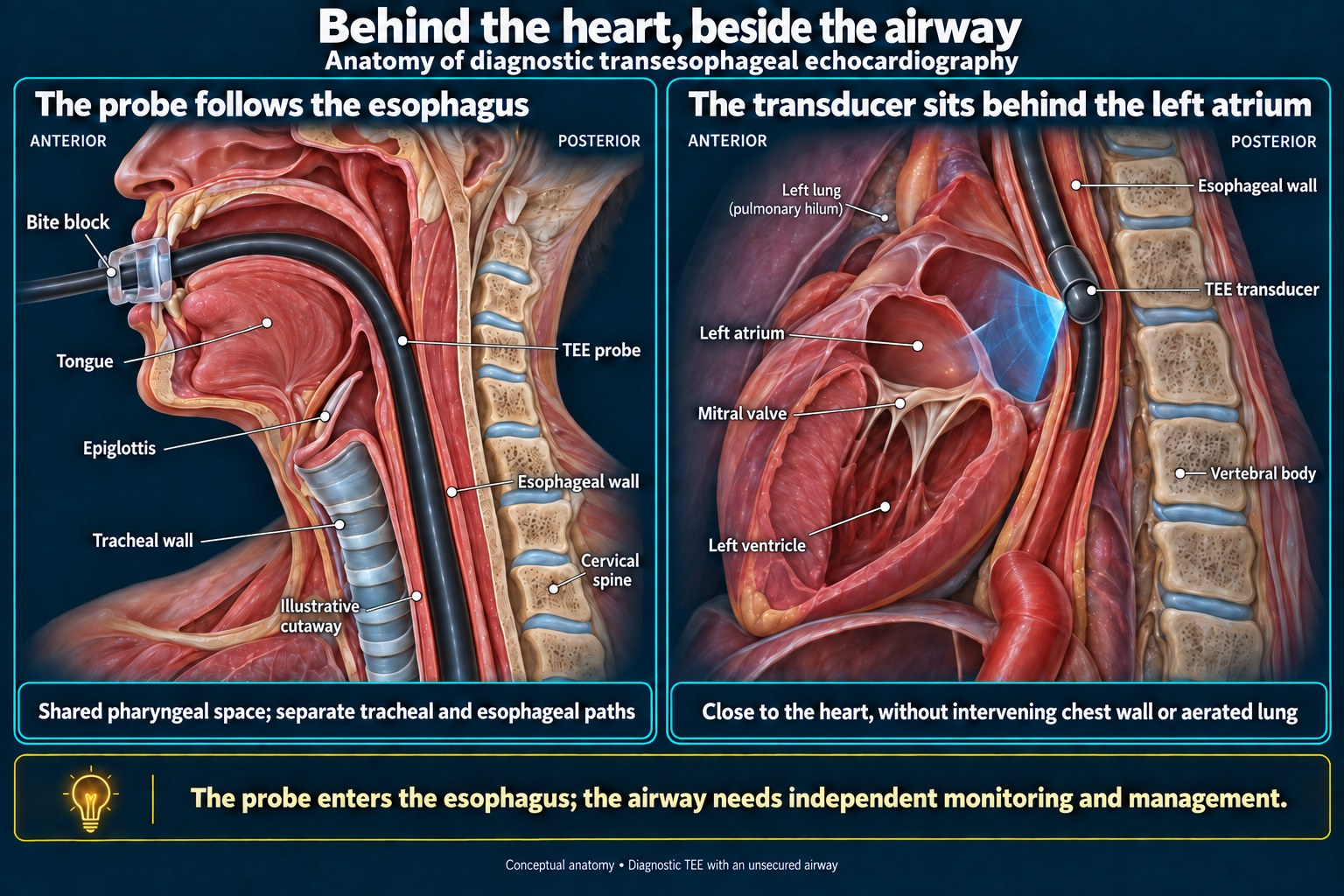
Transesophageal echocardiography puts an ultrasound transducer in the esophagus, directly behind the heart, so the image is not looking through the chest wall or the lungs. For the anesthetist the important sentence is the second one: the probe is large, continuously stimulating, and stays in for the duration.
Key takeaways
- The whole case is one balance: deep enough to tolerate the probe, awake enough to protect an airway the probe is lying against. Nothing satisfies both comfortably.
- Benzocaine spray causes methemoglobinemia at a rate of roughly 1 in 220 to 1 in 1,500 TEEs. This is the most likely drug catastrophe of the case and it is caused by the topicalization, not the sedation. The risk is concentrated almost entirely in inpatients.
- Esophageal perforation occurs in about 0.03–0.09% and carries a mortality near 30%. Most reported perforations followed exams that felt smooth and low-risk.
- Injury declares late. In one review, 11 of 30 perforations were diagnosed more than 24 hours after the procedure.
- Antibiotics are not indicated — TEE is among the nondental procedures that need no endocarditis prophylaxis unless there is active infection at the procedure site, and the American Heart Association has not recommended antibiotics solely to prevent endocarditis for gastrointestinal or genitourinary procedures since 2007. This catches people out because the patient is so often an endocarditis workup.
- Esophageal varices are a weaker contraindication than commonly taught. Pooled data now put post-TEE bleeding in cirrhosis under 1%. Prior esophagectomy is the one most consistently treated as absolute.
What a TEE is, and why it is done from the esophagus
The esophagus sits immediately posterior to the left atrium. A transducer placed there is separated from the heart by millimeters of tissue and no bone or air, which is why TEE resolves structures a transthoracic study cannot — the left atrial appendage, valve vegetations, prosthetic valves, the interatrial septum, the thoracic aorta.
The diagnostic indications you will meet most often
- Before cardioversion — to exclude thrombus in the left atrial appendage.
- Endocarditis workup — vegetations, abscess, prosthetic valve involvement.
- Valve assessment where transthoracic images are inadequate or the question is structural.
- Suspected cardiac source of embolism after stroke or TIA.
A separate use, and a different job: intraoperative TEE during cardiac surgery, where the probe goes in after induction of general anesthesia and stays for the case. The airway is already secured and the sedation problem does not exist — see CABG anesthesia, step by step for that setting. The rest of this page is about the diagnostic study in a sedated patient, which is the harder anesthetic.
This is in the app, with the sedation depth and airway tools — free tier, no card. Get it →
The patients, and the contraindications
A cardiology population — heart failure, valve disease, atrial fibrillation on anticoagulation, suspected endocarditis — often frail. The contraindications the probe cares about are esophageal rather than cardiac.
| Finding | How it is usually treated |
|---|---|
| Prior esophagectomy or esophagogastrectomy | The one most consistently described as absolute1,2 |
| Esophageal stricture, tumor, recent esophageal surgery | Generally absolute or near-absolute; fatal hemorrhage has been reported where the probe disrupted tissue infiltrated by tumor3 |
| Zenker’s diverticulum | Rare but described as a site of life-threatening perforation4 |
| Esophageal varices | Weaker than commonly taught — see below |
| Prominent anterior cervical vertebral osteophytes | Proposed as a relative contraindication; the probe compresses the esophagus against them5 |
| Prior chest radiation, dysphagia history, chronic steroid use, large left atrium | Recognized risk factors for esophageal injury6 |
| Severe atherosclerotic disease | Esophageal perfusion may be compromised; perforation has been reported after entirely smooth insertion6 |
On varices specifically. In a cohort of 401 liver transplant patients, 320 of whom received TEE, postoperative upper GI bleeding occurred in 0.6% of those with varices (1 of 171) versus 2.7% of those without (4 of 149) — not a statistically significant difference, and numerically in the opposite direction from expectation.7 Two 2022 systematic reviews with meta-analysis now support the same conclusion at scale: pooled post-TEE bleeding in patients with cirrhosis was under 1% (about 0.84%), with no significant difference between those with and without varices and no reported perforations or deaths.25,26 A multicenter cohort of 191 cirrhotic patients with documented esophageal varices found no overt gastrointestinal bleeding after TEE and no benefit from routine pre-procedure endoscopy.27 That does not make varices irrelevant, but it does not support treating them as an absolute bar either.
The ASA practice guideline is candid about the state of this evidence: it found insufficient literature to assess whether contraindications exist at all, and its consultants were equivocal on whether anything other than prior esophagectomy or esophagogastrectomy is truly absolute.1 Where oral, esophageal or gastric disease is present, the recommended precautions are to consider other imaging modalities, obtain gastroenterology input, use a smaller probe, limit the examination, avoid unnecessary manipulation, and use the most experienced operator.1
The anesthetic, step by step
Treat the fasting status as a real anesthetic question
Because it is one. The probe sits at the aspiration crossroads — a sedated patient, a suppressed gag, and a large foreign body held in the esophagus for the length of the study. Verify fasting the way you would before any anesthetic, not the way you might before a brief procedure.
Two things make this population worse than the average sedation: heart failure delays gastric emptying, and the exam is frequently added urgently to a day already in progress, which is where the fasting history gets taken casually.
Topicalize — and know what you are spraying
Topical oropharyngeal anesthesia does the work that would otherwise have to be bought with depth of sedation. The more effective the topicalization, the less sedation is needed to tolerate the probe.
Benzocaine spray is the most likely drug catastrophe of this case, and it comes from the topicalization rather than the sedation.
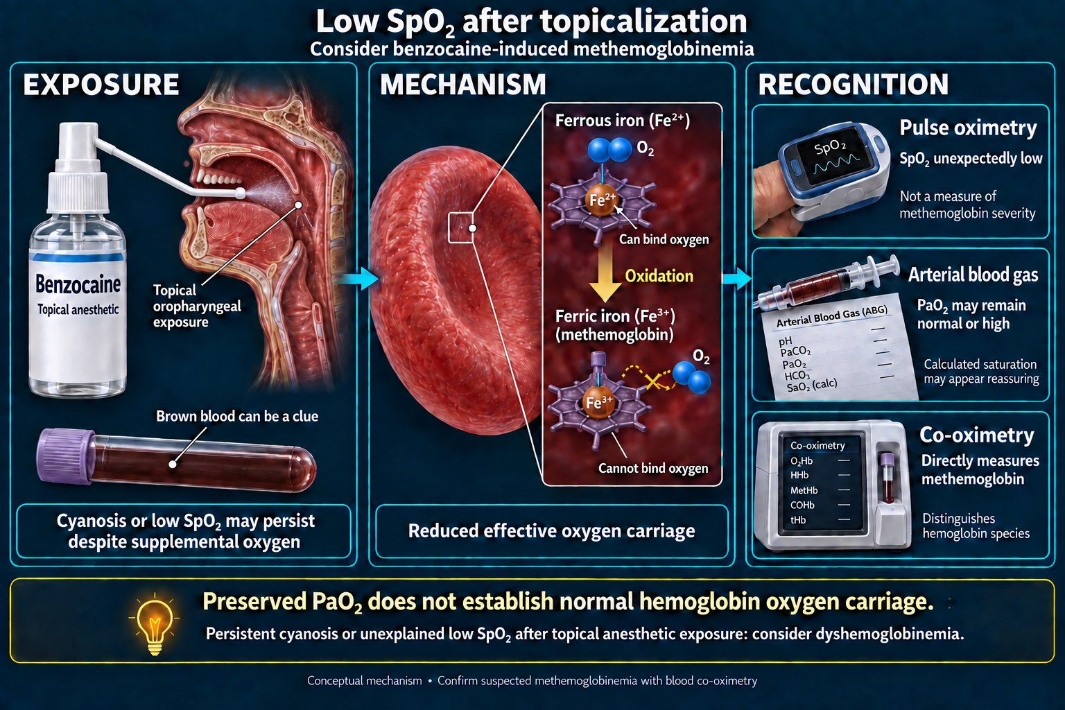
Benzocaine causes acquired methemoglobinemia. Reported incidence during TEE ranges from 0.067% to 0.45% across retrospective series8 — roughly 1 in 220 to 1 in 1,500. The Mayo Clinic series is the largest analysis of incidence, outcomes and predisposing factors: 19 cases in 28,478 transesophageal echocardiograms over 90 months, an incidence of 1 per 1,499 (0.067%), with a mean methemoglobin level of 32%.10 A separate high-volume laboratory reported 5 cases in 4,336 examinations, an incidence of 0.115%.9 All five of those occurred in inpatients; none in the outpatient setting.9 A 10-year case-control study found the same concentration of risk, with 32 of 33 topical-anesthetic methemoglobinemia cases occurring in inpatients — consistent with the added-on, acutely ill patient being the one at risk.32
Individual reported levels vary widely. The Mayo series of 19 cases had a mean of 32%, with a standard deviation of 15%.10 A recent case reached 31.8% in a patient who denied breathlessness,11 and one presented at 69%.12
How it presents, and why it is missed
The signature is a saturation gap: pulse oximetry reads low while arterial oxygen saturation on blood gas reads normal. In one reported case the pulse oximeter showed 75% against an arterial saturation of 99%.12 Another patient developed profound hypoxemia requiring escalation to 100% oxygen by non-rebreather while denying any dyspnea — silent hypoxia with a methemoglobin of 31.8%.11
It is easy to misread as an airway problem in exactly the setting where an airway problem is what you are expecting. A sedated patient with a probe in their esophagus desaturating on the monitor looks like obstruction. Cyanosis unresponsive to oxygen, in a patient who was just sprayed, is the clue.
What to do about it
- Know which agent your unit uses. Benzocaine and Cetacaine (benzocaine, tetracaine and butamben) carry the risk; benzocaine has been removed from some hospital formularies for this reason.8
- Lidocaine is the safer choice. In 3,354 TEEs at one institution using lidocaine over 13 years, there were no cases of clinically evident methemoglobinemia. Its prospective arm of 18 patients did measure a rise, from 0.5% to 0.6% at 60 minutes, which the authors call statistically but not clinically significant — no patient exceeded the normal range or developed symptoms.13
- Treat by threshold, not by the number alone. Give methylene blue to symptomatic patients with methemoglobin above 20%, or to asymptomatic patients above 30%.30
- Know where the methylene blue is, and how to dose it. The dose historically quoted for TEE-related methemoglobinemia is 1–2 mg/kg intravenously, which should reduce methemoglobin significantly in under an hour;21 the patient who presented at 69% received 2 mg/kg with resolution of symptoms.12 The current FDA-approved label is narrower: 1 mg/kg IV over 5–30 minutes, repeated once at 1 mg/kg if methemoglobin remains above 30% or symptoms persist, to a maximum of two doses, with a single 1 mg/kg dose in moderate-to-severe renal impairment; the label states that lower or greater doses are not recommended.29 A treatable emergency is only treatable if the antidote is in the building.
- Methylene blue has its own hazards. It is ineffective and potentially harmful in G6PD deficiency, where it can precipitate hemolysis, and cumulative doses above 7 mg/kg themselves cause cyanosis and hemolytic anemia.30
- Since 2018 the FDA has required a standardized methemoglobinemia warning in the prescribing information of all prescription local anesthetics, and it urged makers of over-the-counter oral benzocaine products to add a methemoglobinemia warning to their labels.14
The mechanism, the reason the pulse oximeter plateaus near 85%, the differential when methylene blue fails, and the G6PD problem are covered on the methemoglobinemia page.
Hold the line between two failures
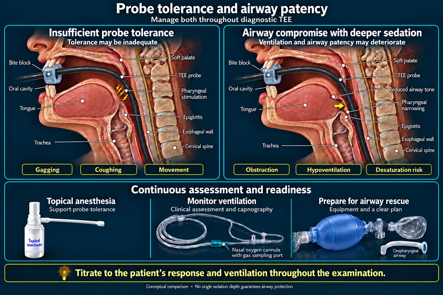
This is the whole case: deep enough to tolerate the probe, awake enough to protect an airway the probe is lying against. The two requirements pull in opposite directions and there is no setting that satisfies both comfortably, which is why this is a harder assignment than its scheduling suggests.
What makes it harder than a comparable endoscopy:
- The stimulus is continuous, not intermittent. There is no quiet interval to let depth drift back up.
- The patient population has less reserve than a general endoscopy list — heart failure, valve disease, frailty.
- The airway is shared with a large device that limits what you can do about obstruction without removing it.
- The operator is looking at a screen, not at the patient. Nobody else in the room is monitoring the airway.
Plan for the cardioversion if it is coming
When cardioversion follows the study in the same encounter, the sedation plan has to include the shock: a brief deepening timed to it, with rhythm drugs and pads already in place. That is a different depth, arriving at a predictable moment — so it is planned rather than improvised.
Practical sequence: probe out first if the study is complete, pads on before the study rather than after, anticoagulation status confirmed and documented, and the deepening timed so the patient is asleep for the shock and not for the fifteen minutes surrounding it.
What to watch for
During the study
Airway obstruction and desaturation with the probe in place is the foreseeable event, and the reason sedation depth is a continuous judgment rather than a bolus. Add benzocaine-induced methemoglobinemia to the differential for any desaturation that does not respond to airway maneuvers or oxygen.
Afterward — and this is the one that is missed
Esophageal injury declares late, and a smooth exam is not reassurance. A systematic review of 35 TEE-induced esophageal perforations from 22 studies found that the majority occurred in cases with a perceived low risk or a smooth examination — so screening for high-risk factors reduces but does not eliminate the event.15 The same review reported a mortality of 28.5%, with shock at the time of detection associated with death.15
Detection is frequently delayed. One review found 11 of 30 perforations diagnosed more than 24 hours after the procedure,6 and in one series both perforations presented more than 48 hours later — on day 4 and day 11.6
Insertion technique is a modifiable factor. A meta-analysis of seven randomized trials found that videolaryngoscope-guided probe insertion reduced overall insertion complications (RR 0.28) and hypopharyngeal mucosal trauma compared with the conventional blind technique, and the ACTACC audit likewise identified insertion as a target for reducing injury.31,20
Post-procedure pain, fever, crepitus, new shortness of breath, pleural effusion or pneumothorax are the findings that should raise it. The discharge instruction matters more than usual here, because the patient will be at home when it declares.
The numbers
| Event | Rate | Source |
|---|---|---|
| Overall complications | Roughly 0.2% in large series; 0.18% with mortality 0.0098% in a multicenter survey of 10,419 exams | Multicenter data16 |
| TEE-related morbidity, intraoperative | 0.2–1.2% | Pooled review17 |
| Esophageal perforation | 0.03–0.09% | Cardiac surgery series5,18 |
| Mortality of perforation once it occurs | 28.5% in systematic review; quoted ranges 10–56% depending on management and time to diagnosis | Sainathan & Andaz15,19 |
| Benzocaine methemoglobinemia | 0.067–0.45% | Retrospective series8,10 |
| Post-TEE bleeding in cirrhosis (pooled) | ~0.84%; no difference by variceal status, no reported perforations or deaths | Meta-analyses25,26 |
| Major complications, prospective national audit | 17 in 22,314 anesthetized examinations — palatal injury or gastro-esophageal disruption | ACTACC UK/Ireland audit20 |
Common minor complications: dysphagia, hoarseness, lip and dental injury.19 Less common but described: vocal cord paralysis, arrhythmia, hypotension, seizure, cardiac arrest.19
Antibiotics
Not indicated. Since its 2007 guideline the American Heart Association has not recommended antibiotics solely to prevent endocarditis for patients undergoing a gastrointestinal or genitourinary tract procedure,22 and its 2021 statement recommended no changes to the 2007 guidelines for preventing viridans group streptococcal endocarditis.23 TEE is among the nondental procedures for which prophylaxis is not required unless there is active infection at the procedure site.24 The 2020 ACC/AHA valvular heart disease guideline is more explicit still, naming TEE among the nondental procedures for which prophylaxis is not recommended even in high-risk patients absent active infection at the site (Class 3: No Benefit).28
This catches people out because the patient population is so often an endocarditis workup — but investigating endocarditis is not an indication to prevent it. The reasoning behind the 2007 change applies here as it does elsewhere: the procedures now covered are those manipulating gingival tissue or the periapical region or perforating oral mucosa, in the highest-risk cardiac patients only.22,24
Frequently asked questions
Do you need antibiotics before a transesophageal echo?
No. TEE is among the nondental procedures for which endocarditis prophylaxis is not required unless there is active infection at the procedure site.24 Since its 2007 guideline the American Heart Association has not recommended antibiotics solely to prevent endocarditis for gastrointestinal or genitourinary tract procedures,22 the 2020 ACC/AHA valvular heart disease guideline names TEE explicitly as not requiring prophylaxis even in high-risk patients (Class 3: No Benefit),28 and the 2021 AHA statement recommended no changes to the 2007 guidelines for preventing viridans group streptococcal endocarditis.23 This is frequently misapplied because the indication for the study is so often suspected endocarditis, but investigating endocarditis is not an indication to prevent it.
What topical anesthetic is used for TEE, and is benzocaine safe?
Benzocaine spray is traditional and carries a real risk of methemoglobinemia — reported at 0.067% to 0.45% during TEE.8,10 Lidocaine appears substantially safer: one institution reported no clinically evident cases across 3,354 TEEs using lidocaine.13 Benzocaine has been removed from some hospital formularies for this reason. Whichever is used, methylene blue should be available.
How is benzocaine methemoglobinemia recognized and treated during a TEE?
By a saturation gap — pulse oximetry reading low while arterial saturation on blood gas is normal. Reported cases include a pulse oximeter reading of 75% against an arterial saturation of 99%,12 and profound hypoxemia in a patient denying any dyspnea.11 Cyanosis unresponsive to supplemental oxygen after topicalization is the clue. Treat symptomatic patients above 20% methemoglobin, or asymptomatic patients above 30%.30 The FDA-labeled dose is methylene blue 1 mg/kg IV over 5–30 minutes, repeated once if needed (maximum two doses); 1–2 mg/kg is the range historically quoted.29,21 Methylene blue is contraindicated in G6PD deficiency.30
How often does TEE perforate the esophagus?
Approximately 0.03% to 0.09%.5,18 Rare, but mortality once it occurs is high — 28.5% in a systematic review of 35 cases, with quoted ranges of 10% to 56% depending on management and time to diagnosis.15,19 The majority of reported perforations followed examinations perceived as low risk or entirely smooth, so screening reduces but does not eliminate the risk.15
Can TEE be done in a patient with esophageal varices?
It is a relative rather than absolute contraindication in most practice. Pooled data from 2022 meta-analyses put post-TEE bleeding in cirrhosis under 1%, with no difference by variceal status and no reported perforations or deaths,25,26 and a multicenter cohort of 191 cirrhotics with documented varices found no overt bleeding and no benefit from routine pre-procedure endoscopy.27 Prior esophagectomy or esophagogastrectomy is the contraindication most consistently treated as absolute.1,2
Why is sedation for a diagnostic TEE difficult?
Because the two requirements conflict. The patient must be deep enough to tolerate a large probe producing continuous stimulation, and awake enough to protect an airway that the probe is lying directly against. Unlike endoscopy there is no quiet interval, the population has limited cardiac reserve, and the operator is watching a screen rather than the patient.
When does esophageal injury from TEE show up?
Often late. One review found 11 of 30 perforations diagnosed more than 24 hours after the procedure, and in one series both perforations presented more than 48 hours later, on day 4 and day 11.6 Post-procedure pain, fever, crepitus, new breathlessness, pleural effusion or pneumothorax should raise it — and the patient will usually be at home by then.
References
- American Society of Anesthesiologists and Society of Cardiovascular Anesthesiologists Task Force on Transesophageal Echocardiography. Practice guidelines for perioperative transesophageal echocardiography. Anesthesiology. 2010;112(5):1084–1096. doi:10.1097/ALN.0b013e3181c51e90. PMID 20418689.
- Samadzadeh Tabrizi N, Gorin AR, Stout PA, et al. Transesophageal echocardiography in patients with esophagectomy: a review. J Cardiothorac Vasc Anesth. 2025;39(1):208–214. doi:10.1053/j.jvca.2023.08.002. PMID 39551697.
- Hilberath JN, Oakes DA, Shernan SK, Bulwer BE, D’Ambra MN, Eltzschig HK. Safety of transesophageal echocardiography. J Am Soc Echocardiogr. 2010;23(11):1115–1127. doi:10.1016/j.echo.2010.08.013. PMID 20864313. Reviewing the Daniel multicenter series, including fatal hemorrhage where probe insertion disrupted tumor-infiltrated esophageal tissue.
- Terryn FX, Stangherlin P, Mansvelt B. Zenker’s diverticulum perforation due to transoesophageal echocardiography. Int J Surg Case Rep. 2020. doi:10.1016/j.ijscr.2020.06.016
- Esophageal perforation with transesophageal echocardiography in an elderly patient with prominent vertebral osteophytes: a case report and review of the literature. CASE (Phila). 2020. PMC7581634. Perforation incidence 0.03–0.09%; osteophytes proposed as relative contraindication.
- Patel KM, Desai RG, Trivedi K, Neuburger PJ, Krishnan S, Potestio CP. Complications of transesophageal echocardiography: a review of injuries, risk factors, and management. J Cardiothorac Vasc Anesth. 2022;36(8 Pt B):3292–3302. doi:10.1053/j.jvca.2022.02.015. PMID 35317955. Reviewing Cote et al. (11 of 30 perforations diagnosed more than 24 hours after the procedure) and Lennon et al. (both perforations presenting on day 4 and day 11).
- Chotalia M, Topiwala U, Iqbal A, et al. Incidence of gastrointestinal bleeding after transesophageal echocardiography use in orthotopic liver transplantation. Transpl Int. 2022;35:10753. doi:10.3389/ti.2022.10753
- Methemoglobinemia due to topical pharyngeal anesthesia during endoscopic procedures. Local Reg Anesth. doi:10.2147/LRA.S12227. Reported benzocaine incidence 0.067–0.45% during TEE.
- Novaro GM, Aronow HD, Militello MA, et al. Benzocaine-induced methemoglobinemia: experience from a high-volume transesophageal echocardiography laboratory. J Am Soc Echocardiogr. 2003;16(2):170–175. doi:10.1067/mje.2003.5. PMID 12574744. Five cases in 4,336 examinations; incidence 0.115% (95% CI 0.037–0.269).
- Kane GC, Hoehn SM, Behrenbeck TR, Mulvagh SL. Benzocaine-induced methemoglobinemia based on the Mayo Clinic experience from 28,478 transesophageal echocardiograms: incidence, outcomes, and predisposing factors. Arch Intern Med. 2007;167(18):1977–1982. doi:10.1001/archinte.167.18.1977. PMID 17923598. Nineteen cases over 90 months; incidence 1 per 1,499 (0.067%, 95% CI 0.040–0.100%); mean methemoglobin 32% ± 15%.
- Easow B, Jiby S, Mathew T, George L, Meek K. Silent hypoxia from benzocaine-induced methemoglobinemia following transesophageal echocardiogram. Cureus. 2025;17(7):e87109. doi:10.7759/cureus.87109. Methemoglobin 31.8% in a patient denying dyspnea.
- A rare side effect of transesophageal echocardiography: methemoglobinemia from topical benzocaine anesthesia. PMID 17074536. Pulse oximetry 75% against arterial saturation 99%; methemoglobin 69%; resolved with methylene blue 2 mg/kg.
- Filipiak-Strzecka D, Kasprzak JD, Wiszniewska M, Walusiak-Skorupa J, Lipiec P. The influence of lidocaine topical anesthesia during transesophageal echocardiography on blood methemoglobin level and risk of methemoglobinemia. Int J Cardiovasc Imaging. 2015. doi:10.1007/s10554-015-0608-z. PMID 25663608. No clinically evident cases in 3,354 TEEs. In the prospective arm of 18 patients, mean methemoglobin rose from 0.5 ± 0.1% to 0.6 ± 0.1% at 60 minutes, a rise the authors describe as statistically but not clinically significant (p = 0.02); no patient exceeded the normal range.
- US Food and Drug Administration. Benzocaine products: risk of methemoglobinemia; standardized warning required on prescription local anesthetics. The announcement is dated 23 May 2018; the FDA page has since been withdrawn, and FDA directs readers to the archived copy: web.archive.org copy of fda.gov.
- Sainathan S, Andaz S. A systematic review of transesophageal echocardiography-induced esophageal perforation. PMID 23834425. Thirty-five cases from 22 studies; mortality 28.5%; majority occurred in perceived low-risk or smooth examinations.
- Jougon JB, Gallon P, MacBride T, et al. Esophageal perforation after transesophageal echocardiography. Eur J Cardiothorac Surg. 1999;16(6):686–687. doi:10.1016/S1010-7940(99)00322-X. PMID 10647846. Citing a multicenter study of 10,419 examinations with complication rate 0.18% and mortality 0.0098%.
- De Luca VM, Cammalleri V, Antonelli G, et al. The other side of the coin: transesophageal echocardiography complications following cardiac surgery and transcatheter structural heart interventions. J Clin Med. 2024;13(15):4291. doi:10.3390/jcm13154291. PMID 39124557. PMC11312835. TEE-related morbidity 0.2–1.2%.
- Singh A, Nanda C, Mehta Y. Transesophageal echocardiography probe-induced esophageal perforation. J Card Crit Care TSS. 2024;8(3):169–171. doi:10.25259/JCCC_57_2023.
- Esophageal perforation during transesophageal echocardiography managed conservatively: a case report with a review of the literature on management. Cureus. PMC11695063. Lists dysphagia, hoarseness, lip and dental injury as common, and vocal cord paralysis, arrhythmia, hypotension, seizure and cardiac arrest as serious.
- Ramalingam G, Choi SW, Agarwal S, et al. Complications related to peri-operative transoesophageal echocardiography — a one-year prospective national audit by the Association of Cardiothoracic Anaesthesia and Critical Care. Anaesthesia. 2020;75(1). doi:10.1111/anae.14734. 22,314 examinations, 17 major complications.
- Iolascon A, Bianchi P, Andolfo I, et al. Recommendations for diagnosis and treatment of methemoglobinemia. Am J Hematol. 2021;96(12):1666–1678. doi:10.1002/ajh.26340. PMID 34467556. PMC9291883. Usual starting dose of methylene blue 1–2 mg/kg intravenously; methemoglobin should fall significantly in less than an hour.
- Wilson W, Taubert KA, Gewitz M, et al. Prevention of infective endocarditis: guidelines from the American Heart Association. Circulation. 2007;116(15):1736–1754. doi:10.1161/CIRCULATIONAHA.106.183095. PMID 17446442. Antibiotics solely to prevent endocarditis are not recommended for genitourinary or gastrointestinal tract procedures; for dental procedures, prophylaxis is reasonable only in the highest-risk cardiac conditions, for procedures that manipulate gingival tissue or the periapical region of teeth or perforate the oral mucosa.
- Wilson WR, Gewitz M, Lockhart PB, et al. Prevention of viridans group streptococcal infective endocarditis: a scientific statement from the American Heart Association. Circulation. 2021;143(20):e963–e978. doi:10.1161/CIR.0000000000000969. PMID 33853363. On review of the evidence since 2007, no recommended changes to the 2007 viridans group streptococcal endocarditis prevention guidelines.
- Wijeysundera DN, Finlayson E. Preoperative evaluation. In: Gropper MA, Eriksson LI, Fleisher LA, et al., eds. Miller’s Anesthesia. 10th ed. Elsevier; 2024:834. Prophylaxis is not required for nondental procedures, transesophageal echocardiography among them, unless there is active infection at the procedure site.
- Odewole M, Sen A, Okoruwa E, et al. Systematic review with meta-analysis: incidence of variceal hemorrhage in patients with cirrhosis undergoing transesophageal echocardiography. Aliment Pharmacol Ther. 2022. Pooled post-TEE bleeding in cirrhosis under 1%; no significant difference by variceal status; no reported perforations or deaths.
- Hui RW, Leung CM. Incidence of gastrointestinal bleeding after transesophageal echocardiography in patients with gastroesophageal varices: a systematic review and meta-analysis. J Am Soc Echocardiogr. 2022. Low pooled bleeding rate with no signal of excess risk attributable to varices.
- Sack JS, Li M, Zucker SD. Bleeding outcomes following transesophageal echocardiography in patients with cirrhosis and esophageal varices. Hepatol Commun. 2021. Multicenter cohort of 191 cirrhotic patients with documented varices; no overt gastrointestinal bleeding after TEE and no benefit from routine pre-procedure endoscopy.
- Otto CM, Nishimura RA, Bonow RO, et al. 2020 ACC/AHA guideline for the management of patients with valvular heart disease: a report of the American College of Cardiology/American Heart Association Joint Committee on Clinical Practice Guidelines. J Am Coll Cardiol. 2021;77(4):e25–e197. doi:10.1016/j.jacc.2020.11.018. Transesophageal echocardiography named among nondental procedures for which endocarditis prophylaxis is not recommended even in high-risk patients absent active infection at the site (Class 3: No Benefit).
- US Food and Drug Administration. Methylene blue injection: prescribing information. Dose 1 mg/kg IV over 5–30 minutes, repeated once at 1 mg/kg if methemoglobin remains above 30% or symptoms persist, maximum two doses; single 1 mg/kg dose in moderate-to-severe renal impairment; lower or greater doses not recommended.
- Twine AJ, Rees DC. Methaemoglobinaemia: from pathophysiology to contemporary clinical management. Br J Haematol. 2026. Treat symptomatic patients above 20% methemoglobin and asymptomatic patients above 30%; methylene blue ineffective and potentially harmful in G6PD deficiency; cumulative doses above 7 mg/kg cause cyanosis and hemolytic anemia.
- Hung KC, Wang WT, Yu CH, et al. Efficacy and safety of videolaryngoscopes for transesophageal echocardiography probe insertion: a trial sequential meta-analysis. PLoS One. 2024. Seven randomized trials; videolaryngoscope-guided insertion reduced overall insertion complications (RR 0.28) and hypopharyngeal mucosal trauma versus the conventional technique.
- Chowdhary S, Bukoye B, Bhansali AM, et al. Risk of topical anesthetic-induced methemoglobinemia: a 10-year retrospective case-control study. JAMA Intern Med. 2013. Thirty-two of 33 topical-anesthetic methemoglobinemia cases occurred in inpatients.
Disclaimer. Reference information for licensed clinicians and students. Not a medical device, and not a substitute for clinical judgment. Verify against institutional protocol and current package inserts.
Drawn from the diagnostic transesophageal echocardiography entry inside Helix Anesthesia, where each statement carries its own source and basis label. See how we source clinical content.
Built for the anesthesia machine, not the desk. It opens in one tap, works with no signal in an OR that has none, and shows the citation on screen beside the number rather than making you go and find it.
Free tier, no card: every calculator, every crisis checklist, the full drug library and glossary, three complete regional blocks, five complete surgical cases, and 15 AI questions a month.
Get Helix Anesthesia How we source itSign up for our newsletter and special offers. New answer pages, improvements to the ones already here, and the occasional offer. Nothing else, and not often.
Local anesthetic and sedation safety