
CABG anesthesia, step by step
An on-pump coronary bypass is not one anesthetic but three transitions with an interval between them — onto bypass, the arrested heart, and separation — each with its own checklist.
Every clinical claim on this page is cited to its source below. How we source clinical content.
The short answer
An on-pump coronary bypass is three transitions rather than one anesthetic: going onto bypass, the arrested interval, and separation. The defining problem is the pre-bypass window, where a myocardium with compromised oxygen supply must survive induction before the operation fixes anything. Heparin 300–400 IU/kg targets an ACT above 480 seconds.
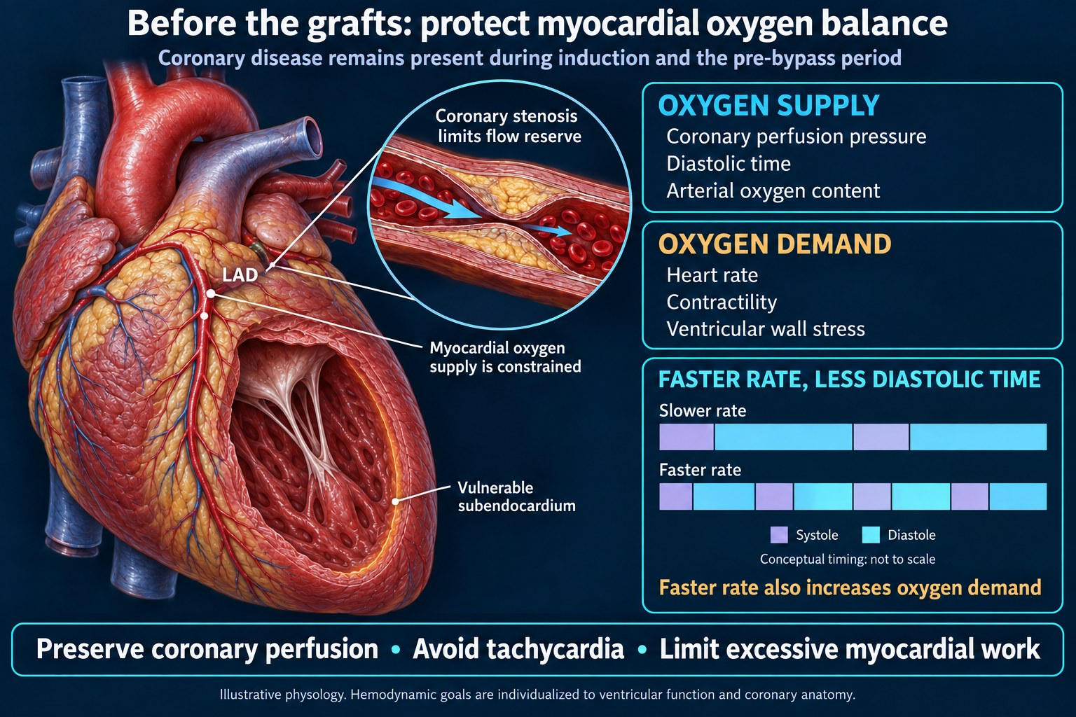
Underneath all of them sits the defining tension: a myocardium whose oxygen supply is already compromised has to survive induction and the pre-bypass period without the ischemia the operation exists to fix.
Key numbers
- Heparin 300–400 IU/kg before cannulation, targeting an ACT above 480 seconds per the STS/SCA/AmSECT guideline. Many institutions accept 400–480.
- Heparin resistance occurs in 4–26% of cases.13 The proposed standardized definition is failure to reach an ACT of 480 seconds after 500 U/kg.5
- Protamine 1 mg per 100 IU of heparin is the traditional ratio, and the guideline direction is downward — dose by titration, and do not exceed 1:1. Recent studies support ratios well below 1:1.20,21,22
- Tranexamic acid roughly halves reoperation for bleeding (1.4% vs 2.8%) and nearly halves blood product use — and raises seizures sevenfold (0.7% vs 0.1%).
- Separation is a list worked in order, not a moment: rhythm, rate, contractility, afterload, preload, with pacing available.
- Emergence usually does not happen in the operating room. The patient leaves sedated and ventilated.
Before induction
Multivessel coronary disease rarely travels alone. Diabetes, carotid and peripheral vascular disease, renal impairment and reduced ventricular function are the usual company, and each changes something. Preoperative antiplatelet and anticoagulant status shapes both the bleeding and the timing question.
Ventricular function on the recent echocardiogram is the single number most worth knowing before induction. It sets how much hypotension the induction can afford and whether inotropic support is a contingency or a plan.
What to establish, and why each matters
| Item | What it changes |
|---|---|
| Ejection fraction and regional wall motion | How much induction hypotension is tolerable; whether inotrope is prepared or merely available |
| Left main or equivalent disease | Narrows the acceptable hemodynamic window further; some centers place lines and start support before induction |
| Antiplatelet agent and last dose | Bleeding risk and transfusion planning. Preoperative aspirin in the ATACAS trial neither reduced thrombotic complications nor increased bleeding1 |
| Renal function | Contrast timing, transfusion threshold, drug clearance |
| Carotid disease | Perfusion pressure targets and, in some centers, cerebral oximetry monitoring |
| Prior heparin exposure, HIT history | Whether heparin is usable at all; alternative anticoagulation requires planning, not improvisation |
| Antithrombin-depleting exposures | Preoperative heparin, liver disease, nephrotic syndrome and sepsis all predispose to heparin resistance2 |
This is in the app, with the full surgical case walkthrough — free tier, no card. Get it →
Lines and monitoring
An arterial line before induction, central venous access, urinary catheter with temperature, and transesophageal echocardiography in most centers. Processed EEG and cerebral oximetry vary by institution.
Five-lead ECG with ST analysis earns its place in this case more than in any other. The thing you are watching for in the pre-bypass window is ischemia, and ST trend is the continuous form of that question.
Positioning is supine with arms tucked and both chest and leg conduit sites prepped. Once draped, access to everything runs through lines placed beforehand — which is the practical argument for placing more than you think you need rather than fewer.
Induction and the pre-bypass window
This is the interval the case is built around. The coronary disease is present, the grafts are not yet, and the hemodynamic goals are the ones that protect a supply-limited myocardium: adequate diastolic pressure for coronary perfusion, a heart rate slow enough to preserve diastolic filling time, and no tachycardia.
Choice of maintenance agent does not drive outcome. The 2021 ACC/AHA/SCAI revascularization guideline notes that although volatile anesthesia may facilitate earlier extubation, the choice of maintenance anesthetic likely does not affect mortality after cardiac surgery.16 The primary evidence is MYRIAD, which randomized 5,400 cardiac surgery patients (64% on-pump CABG) to volatile or total intravenous anesthesia and was stopped for futility: 1-year mortality 2.8% versus 3.0% (RR 0.94, 95% CI 0.69–1.29), with no difference at 30 days.24 A meta-analysis of 89 randomized trials and 14,387 patients also found no mortality difference.25 The choice can be individualized to the patient and the team.
Watch for
- New ST change, a new wall motion abnormality on echo, or hemodynamic deterioration — ischemia in the window where the disease is present and the fix is not.
- Sternotomy, conduit harvest and cannulation each carry their own stimulus and their own hemodynamic consequence. Sternotomy is the largest single stimulus of the pre-bypass period; internal thoracic artery harvest is long and comparatively unstimulating, which is where depth can drift the other way.
- Aortic manipulation. Cannulation and cross-clamping of a calcified aorta are the embolic events of the case, and epiaortic scanning changes the surgical plan in some patients.
Going on bypass
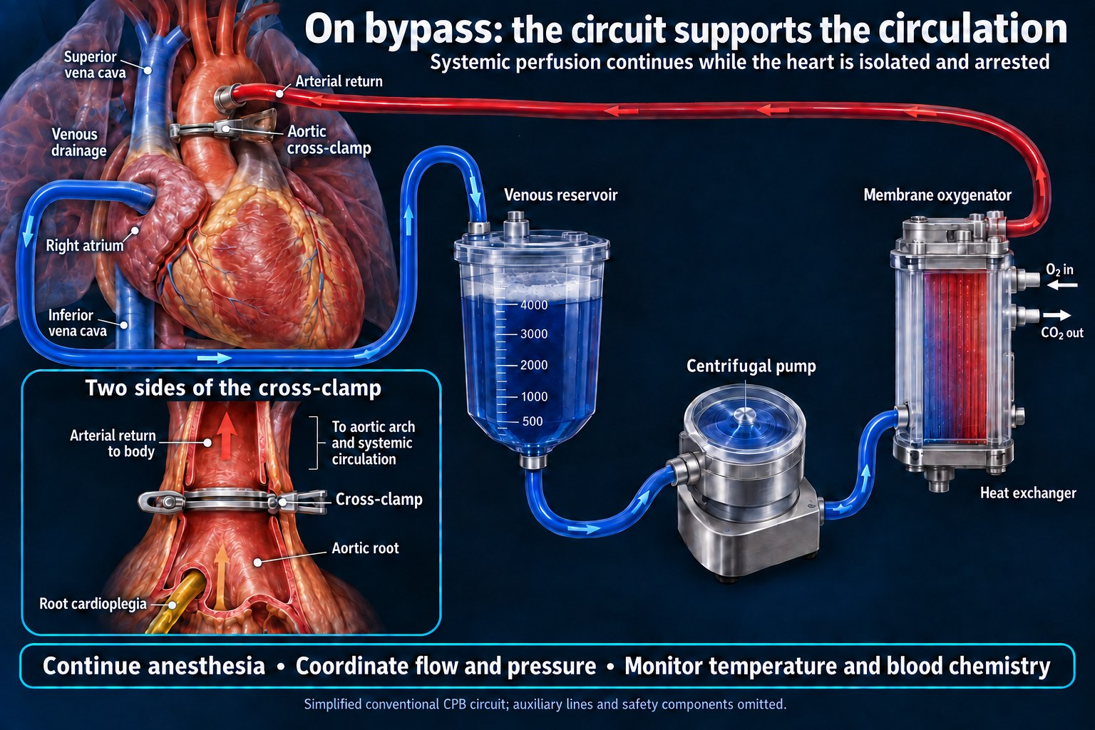
Anticoagulation is given, confirmed by activated clotting time, and announced before cannulation. The announcement is part of the procedure, not courtesy: the surgeon cannot cannulate on an unconfirmed ACT.
The numbers
| Step | Figure | Note |
|---|---|---|
| Heparin bolus | 300–400 IU/kg | Response is heterogeneous between patients; the dose is a starting point, not a guarantee2 |
| ACT before cannulation | >480 seconds | STS/SCA/AmSECT 2018. A margin above 400 seconds, which is where the historical evidence sits. Many institutions accept 400–4803,4 |
| ACT maintained on bypass | >480 seconds | Checked at regular intervals during bypass.4 Routine redosing is the common technique: the STS strategy is one third of the initial heparin bolus at 90 min after the start of bypass, then every 60 min2 |
| Heparin resistance | Incidence 4–26%13 | Proposed standardized definition: ACT below 480 s after 500 U/kg5 |
The 480-second target is softer than it looks, and the guideline says so. It calls the threshold an approximation that may vary with the bias of the instrument being used, and notes that the figure became a “standard of care” on limited evidence.3 Falsely elevated ACT values occur with hypothermia, reduced hemoglobin concentration, hypofibrinogenemia and certain drugs — the first two of which are about to happen — and many ACT tests correlate poorly with heparin level measured as anti-Xa activity.3 Heparin concentration assays and heparinase-modified ACTs exist for exactly this reason, and are used in some centers to distinguish residual heparin effect from other coagulopathy.4,6
If the ACT will not come up
Antithrombin deficiency is the classic cause, since heparin works by potentiating antithrombin rather than acting directly. Additional heparin is the commonest first response; antithrombin concentrate or fresh frozen plasma is the alternative when the problem is a substrate deficiency rather than a dosing one.2,6 Escalating heparin indefinitely has its own costs and does not reliably fix an antithrombin problem.
The arrested interval
The anesthetic does not pause because the heart has. Three things change at once, and all three are easy to lose track of because the usual feedback is gone.
| What changes | Consequence |
|---|---|
| Awareness prevention shifts to the pump | The volatile agent is no longer being delivered by your machine. Whoever is delivering it — the perfusionist via the oxygenator, or you via a TIVA infusion — needs to be explicitly established, not assumed |
| Ventilation stops with the lungs deflated | End-tidal CO₂ disappears as a monitor. Alarms that normally tell you something is wrong now tell you nothing |
| Pressure management becomes a negotiation | Flow belongs to the perfusionist, resistance is yours. Neither of you controls mean arterial pressure alone |
Myocardial protection
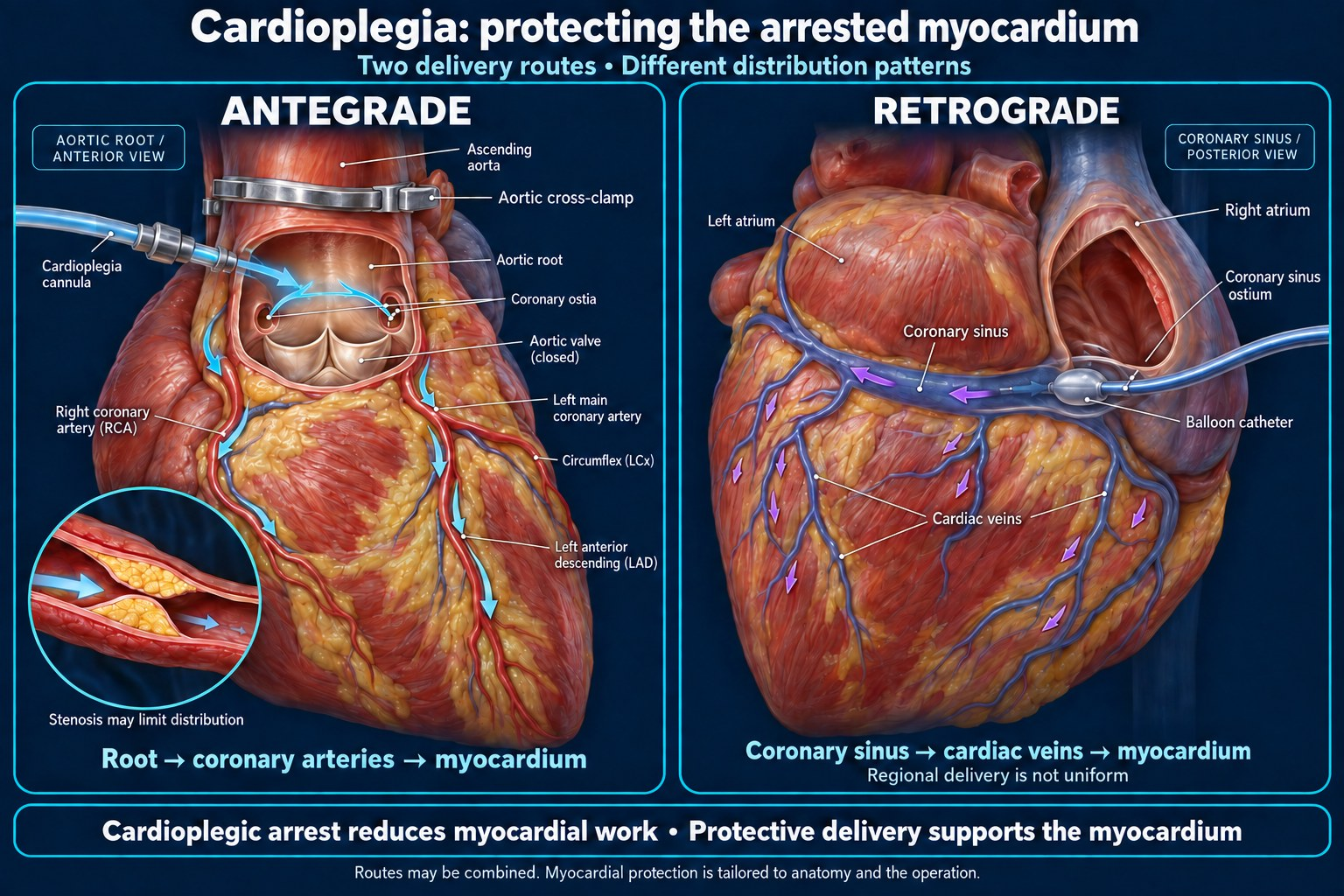
The arrested heart is kept alive by cardioplegia delivered by the surgical team — a hyperkalemic solution that induces diastolic arrest, given after the aorta is cross-clamped.14 Arrest at physiological temperature cuts myocardial oxygen consumption by about 90%, and cooling reduces it further.26 Most centers use blood cardioplegia (Buckberg, del Nido, or microplegia variants) or crystalloid solutions (St Thomas’, histidine-tryptophan-ketoglutarate/Custodiol), delivered antegrade into the aortic root, retrograde via the coronary sinus, or both.14 Blood carries oxygen and dilutes less, and the comparisons favor it: a meta-analysis of 34 trials found less low-output syndrome (OR 0.54) and less CK-MB release with blood cardioplegia, though myocardial infarction and death were similar,28 and a later meta-analysis of 12 studies and 2,866 patients, cited by the 2021 AATS consensus, found lower perioperative myocardial infarction.27 Warm versus cold has shown no clinical-outcome difference,26,27 and single versus multidose no clear winner.14 Distribution is unreliable beyond a tight stenosis and in aortic regurgitation, which is where retrograde or down-graft delivery supplements it, and a myocardial temperature probe serves as a surrogate for adequate delivery.14 Delivery is a surgical responsibility, but the quality of protection during the cross-clamp interval is one of the largest determinants of how the ventricle behaves at separation, so it belongs on the anesthesia professional’s radar — a long cross-clamp time or a difficult cardioplegia delivery is a warning about the separation to come.
Glucose rises on bypass regardless of whether the patient is diabetic, driven by the stress response, hypothermia and catecholamines. Insulin management is protocolized in most units.
Temperature, hematocrit, potassium and acid-base status all move during this interval and all of them need to be where you want them before separation is attempted rather than after it fails.
Scope: this walkthrough describes on-pump, arrested-heart CABG, the most common strategy. Off-pump CABG (OPCAB) avoids the cardioplegic arrest and this separation sequence entirely, trading them for hemodynamic management during cardiac positioning and displacement; on-pump beating-heart is a third variant.23
Separation
Worked as a list, in order, with pacing available. The order matters because each item depends on the one before it — there is no point optimizing contractility in a patient who is in atrial fibrillation.
| Check | Levers | |
|---|---|---|
| 1. Rhythm | Sinus, or paced | Defibrillation, epicardial pacing, antiarrhythmic; correct potassium and magnesium first |
| 2. Rate | Adequate for cardiac output | Pacing is the reliable lever; chronotropes second |
| 3. Contractility | Visual assessment on TEE, not inference | Inotrope, calcium, correct acidosis and hypocalcemia |
| 4. Afterload | Systemic vascular resistance | Vasopressor for vasoplegia, vasodilator if the ventricle is struggling against resistance |
| 5. Preload and flow | Volume handed back gradually while watching the heart fill | Perfusionist returns volume; TEE shows whether the ventricle is tolerating it |
| 6. Rate of ejection | Ventilation resumed, lungs recruited, before flow is fully off | Confirm both lungs are ventilating and the graft territory is not being compressed |
Reperfusion after cross-clamp release is a classic moment for malignant arrhythmia. Have the defibrillator paddles in the field and potassium corrected before the clamp comes off, not after the ventricle fibrillates.
Vasoplegia
If you are recording the vasopressor burden rather than treating it, the norepinephrine-equivalent formulas disagree with each other most on phenylephrine — and none of them can see the inotropes or the pump.
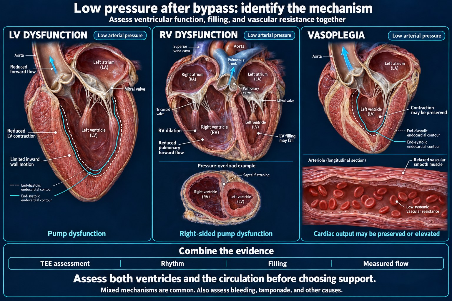
A vasodilated, high-output state after bypass is common — reported in roughly 5–50% of cardiac surgery patients — and is not the same problem as a failing ventricle; treating it with inotrope makes it worse. Distinguishing the two on TEE before escalating is the reason the echo is there. Prior renin-angiotensin-aldosterone inhibition, longer bypass duration, prior cardiac surgery and higher transfusion burden are the recognized risk factors.15,17,18
Management is a mechanism-based escalation rather than more of one agent. A 2021 expert consensus strongly recommends norepinephrine and/or vasopressin to restore and maintain perfusion pressure, though it cannot recommend either over the other with respect to ischemic complications.29 Recent management algorithms place norepinephrine first, with early addition of vasopressin, and angiotensin II as the next step for escalating support;15,17,18 the consensus, by contrast, found insufficient evidence to make any recommendation on angiotensin II in cardiac surgical patients.29
The consensus weakly recommends considering early addition of a second vasopressor when one agent cannot restore vascular tone, and considering vasopressin as the first-line vasopressor, or adding it to norepinephrine, in patients with pulmonary hypertension or right-sided heart dysfunction.29 It makes the same weak recommendation to prevent atrial arrhythmias (moderate-quality evidence, 95.2% agreement); one panelist dissented, noting it could be read as advising vasopressin first-line for every cardiac surgical patient.29 Among the evidence it cites, a trial of first-line vasopressin versus norepinephrine in vasoplegic shock after cardiac surgery found vasopressin superior on a combined outcome of 30-day mortality and severe complications, with less acute kidney injury and atrial fibrillation and shorter ICU and hospital stays.29
Methylene blue (a guanylate cyclase inhibitor), hydroxocobalamin and corticosteroids are adjuncts reserved for refractory cases, chosen by mechanism and adverse-effect profile.15,17,18 The same consensus strongly recommends against using methylene blue for anything other than rescue therapy, and against dopamine for post-cardiac surgery vasoplegic shock, both on low-quality evidence with full panel agreement.29
The failure mode to have thought about in advance
Failure to separate: a ventricle that cannot carry the circulation despite pacing and inotropes, escalating toward mechanical support. The decision point is not a moment of realization at the table — it is a threshold agreed with the surgeon before the attempt, so that going back on bypass is a planned step rather than a defeat. Right ventricular failure is a distinct and often under-recognized version of this, and it responds to different interventions than left ventricular failure.
After bypass
Protamine
Given slowly, with the surgical field watched. The traditional dose is 1 mg of protamine per 100 IU of heparin, and the guideline direction has been downward — dose should be based on titration to existing heparin concentration and should not exceed a 1:1 ratio, because excess protamine is itself an anticoagulant and contributes to bleeding.6 Recent studies support ratios well below 1:1: a population pharmacokinetic–pharmacodynamic model predicted that a ratio of about 0.6:1 (0.625:1) would fully reverse heparin in 95% of patients,20 an observational cohort found 0.5:1 sufficient to neutralize residual heparin in most patients,21 and a randomized trial found a fixed 250 mg dose comparable to weight-based 1:1 dosing on post-reversal ACT and chest-tube output while using substantially less protamine.22 A low-dose protamine infusion of 25 mg/h for six hours after neutralization has been reported to prevent heparin rebound, measured by thrombin clotting time and anti-factor Xa concentration — though in the same study the accompanying reduction in postoperative bleeding was not clinically relevant.6 The STS/SCA/AmSECT guideline gives it a class IIb recommendation (level of evidence C): a low-dose infusion of 25 mg/h for as long as 6 hours after the end of bypass may be considered as part of a multimodality blood conservation program, based on a 300-patient randomized trial in which it abolished heparin rebound and modestly reduced chest-tube blood loss, though not transfusion.3 Note the six-hour limit. It is not incidental: the argument against exceeding a 1:1 ratio is that surplus protamine anticoagulates, and an infusion without an endpoint is a way of exceeding it slowly.
Protamine reactions are conventionally described in three types, and they are not variations of the same event:
| Type | What happens | Response |
|---|---|---|
| I | Hypotension from rapid administration — histamine-mediated, dose-rate dependent | Slow or stop the infusion; usually resolves |
| II | Anaphylactic or anaphylactoid reaction | Standard anaphylaxis management. Prior protamine exposure, NPH insulin use, fish allergy and prior vasectomy are the described risk factors |
| III | Catastrophic pulmonary vasoconstriction with acute pulmonary hypertension and right heart failure | The one that kills. Stop protamine, support the right ventricle, and consider that reinstitution of bypass with adequate anticoagulation may be lifesaving.3 The reaction is nitric oxide/cGMP-dependent and endothelium-mediated, which the guideline says suggests methylene blue may be the treatment of choice, though high-level evidence to support it is lacking3,19 |
Antifibrinolytics — and the trade-off worth knowing
Intravenous tranexamic acid is standard, and the Society of Thoracic Surgeons guideline gives it a class IA recommendation for reducing bleeding in cardiac surgery.7 The evidence behind it is unusually good, and so is the evidence for its principal harm.
ATACAS randomized 4,631 patients undergoing coronary artery surgery to tranexamic acid or placebo:1
| Outcome | TXA | Placebo | |
|---|---|---|---|
| Death or thrombotic complications at 30 days | 16.7% | 18.1% | RR 0.92 (0.81–1.05), not significant |
| Reoperation for hemorrhage or tamponade | 1.4% | 2.8% | p = 0.001 |
| Total blood product units transfused | 4,331 | 7,994 | p < 0.001 |
| Postoperative seizures | 0.7% | 0.1% | RR 7.62 (1.77–68.71), p = 0.002 |
Two details that matter more than the headline. The tranexamic acid dose was reduced mid-trial from 100 mg/kg to 50 mg/kg because of accumulating seizure reports, and outcomes including seizure risk did not differ between the two doses in that underpowered comparison.1,8 Separately, a meta-analysis of 16 studies enrolling 45,235 patients found an odds ratio of 4.13 (95% CI 2.59–6.57) for seizures with tranexamic acid in adult cardiac surgery.9
At one year, death or severe disability was 3.8% with tranexamic acid and 4.4% with placebo — no significant difference.10 The drug reduces bleeding without a measurable cost in death or disability, and with a small absolute increase in a frightening complication. That is the trade, stated honestly.
A dose-response relationship for seizures is consistently described, with risk concentrated above roughly 2 g/day,12 and lower-dose intravenous regimens have been reported as equally effective for transfusion reduction without the seizure signal.11 The attempt to avoid the problem by giving tranexamic acid topically was tested in the DEPOSITION trial across 3,242 patients in six countries: topical administration produced an 8.3% absolute increase in transfusion without reducing seizures, and the trial was stopped early for safety, leaving it underpowered for the seizure comparison it was designed to answer.7
Bleeding after protamine
Surgical, coagulopathic, or both — and the distinction is made by looking, not by guessing. Transfusion decisions are made against laboratory and viscoelastic data rather than by sight of the field. Re-exploration for bleeding is a known endpoint of the case, not a surprise, and blood should be in the room rather than in the building.
Emergence
Usually not in the operating room. The patient leaves sedated and ventilated for intensive care, where warming completes, bleeding declares itself or does not, and extubation follows a protocol measured in hours. Fast-track extubation pathways vary by institution.
The handover is part of the anesthetic. Total heparin given, total protamine given, ACT at closure, products transfused, inotrope and vasopressor doses with the reason for each, the ventricular function seen on separation, and the surgeon’s assessment of graft flow — all of it is information the intensive care team cannot reconstruct.
Frequently asked questions
How much heparin is given before cardiopulmonary bypass?
300–400 IU/kg is the standard initial bolus, though individual response is heterogeneous and additional doses are frequently required.2 Some centers start at 400 IU/kg or higher.
What ACT is required before going on bypass?
The 2018 STS/SCA/AmSECT guideline considers it reasonable to maintain an activated clotting time above 480 seconds during bypass, described as a margin of safety above 400 seconds.3,4 Practice varies — many institutions target 400–480 seconds, and the guideline itself describes the threshold as an approximation that may vary with the bias of the instrument used, adopted as a standard of care on limited evidence.3
What is heparin resistance and how common is it?
Failure to achieve the target ACT despite an adequate heparin dose.2 Reported incidence is 4–26%, varying with the bolus used and the target chosen.13 An ISTH subcommittee has proposed standardizing the definition as failure to reach an ACT of 480 seconds or more after 500 U/kg.5 Antithrombin deficiency is the classic cause, and antithrombin concentrate or fresh frozen plasma is the treatment when additional heparin does not work.
How much protamine is given to reverse heparin?
The traditional ratio is 1 mg of protamine per 100 IU of heparin, given slowly. Guideline direction has moved toward lower doses titrated to residual heparin concentration rather than to the total heparin given, and the dose should not exceed a 1:1 ratio — excess protamine is itself an anticoagulant.6 Recent studies support ratios well below 1:1, including roughly 0.5–0.6:1 and fixed low-dose strategies.20,21,22
What is a protamine reaction?
Three types are conventionally described: rate-related hypotension from histamine release, anaphylactic or anaphylactoid reaction, and catastrophic pulmonary vasoconstriction with acute right heart failure. For the third, reinstitution of cardiopulmonary bypass with adequate anticoagulation may be lifesaving. The reaction is nitric oxide/cGMP-mediated, and the guideline suggests methylene blue may be the treatment of choice, though high-level evidence is lacking.3,19
Does tranexamic acid cause seizures in cardiac surgery?
Yes, and the effect is real but small in absolute terms. In ATACAS, seizures occurred in 0.7% of the tranexamic acid group versus 0.1% of the placebo group.1 A meta-analysis of 45,235 patients found an odds ratio of 4.13 for seizures.9 The effect appears dose-dependent, concentrated above roughly 2 g/day.12 Against that, tranexamic acid halved reoperation for hemorrhage and nearly halved blood product use, with no difference in death or disability at one year.1,10
What is the order for separating from cardiopulmonary bypass?
Rhythm, rate, contractility, afterload, then preload and flow handed back, with pacing available and ventilation resumed. The order matters because each item depends on the one before it. Reperfusion after cross-clamp release is a recognized moment for malignant arrhythmia.
Are CABG patients extubated in the operating room?
Usually not. The patient typically leaves sedated and ventilated for intensive care, where rewarming completes and bleeding declares itself. Fast-track extubation pathways exist and vary by institution.
References
- Myles PS, Smith JA, Forbes A, et al; ATACAS Investigators of the ANZCA Clinical Trials Network. Tranexamic acid in patients undergoing coronary-artery surgery. N Engl J Med. 2017;376(2):136–148. doi:10.1056/NEJMoa1606424. 4,631 patients in the tranexamic acid comparison (2,311 versus 2,320). Aspirin arm reported separately: Myles PS, et al. Stopping vs. continuing aspirin before coronary artery surgery. N Engl J Med. 2016;374:728–737.
- Cartwright B, Mundell N. Anticoagulation for cardiopulmonary bypass: part one. BJA Educ. 2023;23(3):110–116. doi:10.1016/j.bjae.2022.12.003. PMID 36844440. PMC9947996. Heparin dosing, heparin resistance definition and causes.
- Shore-Lesserson L, Baker RA, Ferraris VA, et al. The Society of Thoracic Surgeons, the Society of Cardiovascular Anesthesiologists, and the American Society of ExtraCorporeal Technology: clinical practice guidelines — anticoagulation during cardiopulmonary bypass. Ann Thorac Surg. 2018;105(2):650–662. doi:10.1016/j.athoracsur.2017.09.061. PMID 29362176.
- Cardiopulmonary bypass: anticoagulation and antagonism. OpenAnesthesia. openanesthesia.org/keywords/cardiopulmonary-bypass-anticoagulation-and-antagonism. Accessed August 2026. ACT targets, factors influencing ACT, heparinase assays.
- Levy JH, Sniecinski RM, Maier CL, et al. Finding a common definition of heparin resistance in adult cardiac surgery: communication from the ISTH SSC subcommittee on perioperative and critical care thrombosis and hemostasis. J Thromb Haemost. 2024;22(4):1249–1257. doi:10.1016/j.jtha.2024.01.001. PMID 38215912. Proposes ACT below 480 s after 500 U/kg as the standardized definition.
- Boer C, Meesters MI, Veerhoek D, et al. Anticoagulant and side-effects of protamine in cardiac surgery: a narrative review. Br J Anaesth. 2018;120(5):914–927. doi:10.1016/j.bja.2018.01.023. PMID 29661409.
- Lamy A, Sirota DA, Jacques F, Poostizadeh A, Noiseux N, Efremov S, et al. Topical versus intravenous tranexamic acid in patients undergoing cardiac surgery: the DEPOSITION randomized controlled trial. Circulation. 2024. doi:10.1161/CIRCULATIONAHA.124.069606. 3,242 patients, 16 hospitals, 6 countries; stopped early; topical produced an 8.3% absolute increase in transfusion without reducing seizures. Cites the STS class IA recommendation for intravenous tranexamic acid.
- Roebker J, Deghan S. Tranexamic acid reduces bleeding risk during coronary artery surgery: the ATACAS trial. 2 Minute Medicine, 11 January 2017. 2minutemedicine.com. Trial summary noting the mid-trial dose reduction from 100 mg/kg to 50 mg/kg and the underpowered dose comparison.
- Takagi H, Ando T, Umemoto T. Seizures associated with tranexamic acid for cardiac surgery: a meta-analysis of randomized and non-randomized studies. J Cardiovasc Surg (Torino). 2017. PMID 28263046. Sixteen studies, 45,235 patients; odds ratio 4.13 (95% CI 2.59–6.57), P < 0.00001.
- Myles PS, Smith JA, Forbes A, et al. Tranexamic acid in coronary artery surgery: one-year results of the ATACAS trial. J Thorac Cardiovasc Surg. 2019;157(2):644–652. PMID 30459103. Death or disability at one year 3.8% versus 4.4%.
- Guo J, Gao X, Ma Y, et al. Different dose regimes and administration methods of tranexamic acid in cardiac surgery: a meta-analysis of randomized trials. BMC Anesthesiol. 2019;19:129. doi:10.1186/s12871-019-0772-0. Forty-nine studies, 10,591 patients.
- International Society of Blood Transfusion. Antifibrinolytic agent — tranexamic acid. Patient blood management resources, Clinical Transfusion Working Party. Zolfaghari S, van Kraaij M; revised by Zolfaghari S, Relke N. Updated January 2026. isbtweb.org/isbt-working-parties/clinical-transfusion/resources/patient-blood-management-resources/3-tranexamic-acid.html. Notes dose-dependent seizure risk above 2 g/day and summarizes the OPTIMAL trial.
- Chen Y, Phoon PHY, Hwang NC. Heparin resistance during cardiopulmonary bypass in adult cardiac surgery. J Cardiothorac Vasc Anesth. 2022;36(11):4150–4160. doi:10.1053/j.jvca.2022.06.021. PMID 35927191. PMC9225936. Incidence 4–26%; survey data on practice variation.
- Sabe SA, Harris DD, Broadwin M, Sellke FW. Cardioprotection in cardiovascular surgery. Basic Res Cardiol. 2024;119(4):545–568. doi:10.1007/s00395-024-01062-0. Cardioplegia strategies, blood vs crystalloid, antegrade/retrograde delivery.
- Chatterjee S, Ortoleva J, Arora RC, et al. Management algorithm for vasoplegic shock after cardiac surgery: an interdisciplinary collaboration. Ann Thorac Surg. 2026. doi:10.1016/j.athoracsur.2026.06.013. Incidence 5–50%; risk factors; norepinephrine, early vasopressin, then angiotensin II; adjuncts for refractory cases.
- Lawton JS, Tamis-Holland JE, Bangalore S, et al. 2021 ACC/AHA/SCAI guideline for coronary artery revascularization: a report of the American College of Cardiology/American Heart Association Joint Committee on Clinical Practice Guidelines. J Am Coll Cardiol. 2022. doi:10.1016/j.jacc.2021.09.006. PMID 34895950. Anesthetic maintenance technique and mortality.
- Datt V, Wadhhwa R, Sharma V, et al. Vasoplegic syndrome after cardiovascular surgery: a review of pathophysiology and outcome-oriented therapeutic management. J Card Surg. 2021;36(10):3749–3760. doi:10.1111/jocs.15805.
- Ltaief Z, Ben-Hamouda N, Rancati V, et al. Vasoplegic syndrome after cardiopulmonary bypass in cardiovascular surgery: pathophysiology and management in critical care. J Clin Med. 2022;11(21):6407. doi:10.3390/jcm11216407. PMID 36362635.
- Viaro F, Dalio MB, Evora PR. Catastrophic cardiovascular adverse reactions to protamine are nitric oxide/cyclic guanosine monophosphate dependent and endothelium mediated: should methylene blue be the treatment of choice? Chest. 2002;122(3):1061–1066. doi:10.1378/chest.122.3.1061. PMID 12226053.
- Lanoiselée J, Gibert A, Gouin-Thibault I, et al. Optimising protamine dosing for heparin reversal after cardiopulmonary bypass: a population pharmacokinetic–pharmacodynamic study. Br J Anaesth. 2026;136(3):847–855. PMID 41638976. 68 patients; simulations predicted a 0.625:1 ratio would fully reverse heparin in 95%.
- Taneja R, Szoke DJ, Hynes Z, Jones PM. Minimum protamine dose required to neutralize heparin in cardiac surgery: a single-centre, prospective, observational cohort study. Can J Anaesth. 2023;70(2):219–227. doi:10.1007/s12630-022-02364-4. PMID 36471142. A 0.5:1 ratio neutralized residual anti-IIa and anti-Xa activity in most patients.
- Jain P, Silva-De Las Salas A, Bedi K, et al. Protamine dosing for heparin reversal after cardiopulmonary bypass: a double-blinded prospective randomized control trial comparing two strategies. Anesthesiology. 2025;142:98–106. doi:10.1097/ALN.0000000000005256. PMID 39388600. Fixed 250 mg dose vs weight-based 1:1.
- Phothikun A, Nawarawong W, Tantraworasin A, Phinyo P, Tepsuwan T. The outcomes of three different techniques of coronary artery bypass grafting: on-pump arrested heart, on-pump beating heart, and off-pump. PLoS One. 2023;18(5):e0286510. doi:10.1371/journal.pone.0286510. PMID 37256890. PMC10231783.
- Landoni G, Lomivorotov VV, Nigro Neto C, et al. Volatile anesthetics versus total intravenous anesthesia for cardiac surgery. N Engl J Med. 2019;380(13):1214–1225. doi:10.1056/NEJMoa1816476. The MYRIAD trial.
- Jiao XF, Lin XM, Ni XF, et al. Volatile anesthetics versus total intravenous anesthesia in patients undergoing coronary artery bypass grafting: an updated meta-analysis and trial sequential analysis of randomized controlled trials. PLoS One. 2019;14(10):e0224562. doi:10.1371/journal.pone.0224562. PMID 31661512.
- Pufulete M, Reeves B, Rogers C, et al. Warm versus cold blood, and any blood versus crystalloid, cardioplegia in adults undergoing coronary artery bypass grafting surgery with cardiopulmonary bypass. Cochrane Database Syst Rev. 2013. doi:10.1002/14651858.CD010650. Carries Buckberg’s figure of about 90% less myocardial oxygen consumption at arrest.
- Bakaeen FG, Gaudino M, Whitman G, et al. 2021: The American Association for Thoracic Surgery expert consensus document: coronary artery bypass grafting in patients with ischemic cardiomyopathy and heart failure. J Thorac Cardiovasc Surg. 2021;162(3):829–850.e1. doi:10.1016/j.jtcvs.2021.04.052.
- Guru V, Omura J, Alghamdi AA, Weisel R, Fremes SE. Is blood superior to crystalloid cardioplegia? A meta-analysis of randomized clinical trials. Circulation. 2006;114(1 Suppl):I331–I338. doi:10.1161/CIRCULATIONAHA.105.001644. PMID 16820596.
- Guarracino F, Habicher M, Treskatsch S, et al. Vasopressor therapy in cardiac surgery — an experts’ consensus statement. J Cardiothorac Vasc Anesth. 2021;35(4):1018–1029. doi:10.1053/j.jvca.2020.11.032. PMID 33334651. GRADE/Delphi consensus: norepinephrine and/or vasopressin (strong); vasopressin in pulmonary hypertension or right-sided dysfunction, and to prevent atrial arrhythmias (weak); methylene blue only as rescue and no dopamine (strong); angiotensin II (insufficient evidence).
Disclaimer. Reference information for licensed clinicians and students. Not a medical device, and not a substitute for clinical judgment. Anticoagulation targets, protamine dosing and antifibrinolytic regimens vary substantially between institutions and testing platforms. Verify against institutional protocol and current package inserts.
Drawn from the coronary artery bypass entry inside Helix Anesthesia, where each statement carries its own source and basis label, and approaches are listed without ranking because plans vary by institution, surgeon and patient. See how we source clinical content.
Built for the anesthesia machine, not the desk. It opens in one tap, works with no signal in an OR that has none, and shows the citation on screen beside the number rather than making you go and find it.
Free tier, no card: every calculator, every crisis checklist, the full drug library and glossary, three complete regional blocks, five complete surgical cases, and 15 AI questions a month.
Get Helix Anesthesia How we source itSign up for our newsletter and special offers. New answer pages, improvements to the ones already here, and the occasional offer. Nothing else, and not often.