
Anesthetic considerations for valvular heart disease
A complete perioperative guide for anesthesia providers — lesion-specific hemodynamic goals, aortic stenosis, neuraxial safety, and the timing of valve intervention before noncardiac surgery.
Every clinical claim on this page is cited to its source below. How we source clinical content.
The short answer
There is no single “valvular” anesthetic — the lesion, its severity, and whether the patient is symptomatic decide the plan. Stenotic lesions are far less forgiving than regurgitant ones. Severe aortic stenosis is the highest-risk lesion and the one to identify before induction: maintain preload and sinus rhythm with a normal heart rate, avoid tachycardia and hypotension, and treat hypotension with phenylephrine or norepinephrine. Patients with severe aortic or mitral stenosis who meet criteria for valve intervention should be evaluated for it before elective elevated-risk surgery.
Key takeaways
- Lesion + severity + symptoms drive the plan. Symptomatic severe aortic stenosis (AS) carries the highest perioperative mortality; regurgitant lesions with preserved left ventricular (LV) function are generally better tolerated than stenotic ones.1,2
- Aortic stenosis is “full, slow, tight, and forward,” with “slow” meaning avoid tachycardia and keep a normal heart rate, not a deliberately slow one. Maintain preload and sinus rhythm with a normal heart rate, avoid both tachycardia and hypotension, preserve systemic vascular resistance and coronary perfusion pressure, and support blood pressure with phenylephrine or norepinephrine.2
- Severe AS meeting criteria for intervention should be evaluated for aortic valve replacement (AVR) before elective noncardiac surgery, which reduces perioperative risk. Balloon aortic valvuloplasty is a possible bridge before urgent surgery.1,10,11
- Neuraxial anesthesia is not absolutely contraindicated in AS, but rapid sympathectomy must be avoided; slow-titrated epidural or carefully dosed technique with invasive pressure monitoring is preferred.2,7,8
- Endocarditis prophylaxis is now reserved for the highest-risk patients only — prosthetic valves or prosthetic repair material, prior endocarditis, certain congenital lesions, and transplant valvulopathy — and only before dental procedures involving gingival or periapical manipulation.2,12,15
- Do not confuse fixed AS with dynamic LV outflow obstruction (HCM). In HCM, positive inotropic agents, tachycardia, and reduced preload are harmful and should be avoided. The two share their preload, rate, and afterload goals, with afterload maintained in both; they differ on contractility, and hypotension in HCM is treated with alpha-agonists, such as phenylephrine or vasopressin, rather than beta-agonists.1,14
Why the lesion matters more than the label
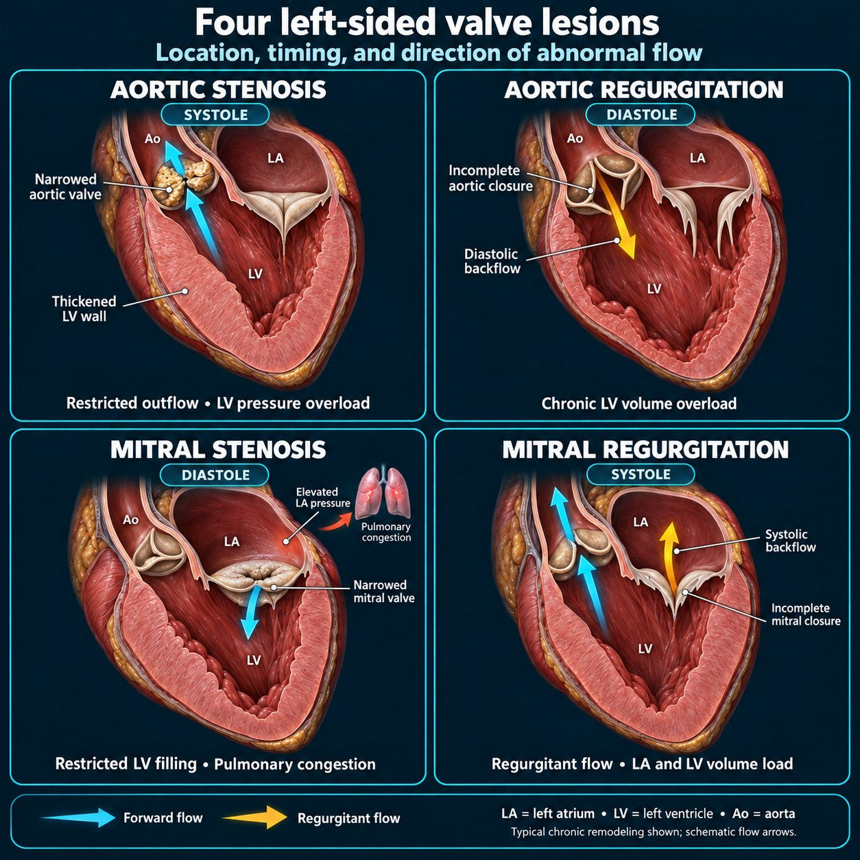
Valvular heart disease is common, particularly in older adults, and is frequently discovered during preoperative assessment.1 “The patient has valve disease” is not enough to plan an anesthetic. The four common left-sided lesions impose opposite loading requirements: a stenotic valve needs a full ventricle, unhurried filling, and preserved perfusion pressure, whereas a regurgitant valve tolerates — and often benefits from — vasodilation and avoidance of bradycardia.2
Two principles cut across all of them. First, echocardiography within 12 months is recommended before elevated-risk elective surgery in any patient with suspected moderate or severe valve disease, to define severity, LV function, other lesions, and pulmonary pressures.1 Second, when a lesion is severe enough to meet independent criteria for valve intervention, that intervention should generally be considered before elective elevated-risk surgery rather than after.1,2
Hemodynamic goals at a glance
The single most useful bedside framework is a lesion-by-lesion account of preload, rate and rhythm, afterload, and the pitfalls to avoid.2
| Lesion | Preload | Rate & rhythm | Afterload (SVR) | Avoid |
|---|---|---|---|---|
| Aortic stenosis | Maintain / full | Sinus rhythm with normal heart rate | Maintain — preserves coronary perfusion of the hypertrophied ventricle | Tachycardia and systemic hypotension (decreased coronary perfusion pressure)2 |
| Aortic regurgitation | Maintain | Avoid bradycardia (increases total diastolic time) | Reduce — lower SVR is favorable in regurgitant valve lesions | Bradycardia, increased afterload2 |
| Mitral stenosis | Maintain, but avoid overload / pulmonary edema | Avoid tachycardia; sinus rhythm; prolong diastolic filling | Maintain | Tachycardia, new atrial fibrillation, hypoxia/hypercarbia (worsen pulmonary hypertension)1,2 |
| Mitral regurgitation | Maintain | Avoid bradycardia; normal-to-slightly-fast rate | Reduce — lower SVR favors forward over regurgitant flow | Bradycardia, increased afterload2 |
This is in the app, with the calculators and the drug library — free tier, no card. Get it →
Aortic stenosis — the highest-risk lesion
Severe AS produces a fixed obstruction to LV outflow, driving compensatory concentric hypertrophy, which reduces myocardial compliance as well as coronary flow reserve.11 Diastolic dysfunction and an increase in LV end-diastolic pressure follow, and the atrial kick may contribute as much as 40% of total cardiac output.17
Grading severity
Transthoracic echocardiography is the reference method, using peak transvalvular velocity, mean pressure gradient, and aortic valve area (AVA) by the continuity equation.4,5
| Grade | Peak velocity | Mean gradient | Valve area |
|---|---|---|---|
| Mild2,5 | 2.0–2.9 m/s | <20 mmHg | >1.5 cm² |
| Moderate2,5 | 3.0–3.9 m/s | 20–39 mmHg | 1.0–1.5 cm² |
| Severe2,5 | ≥4.0 m/s | ≥40 mmHg | <1.0 cm² (≤0.6 cm²/m² indexed) |
| Very severe2 | ≥5.0 m/s | ≥60 mmHg | — |
Velocity and gradient bands follow the ACC/AHA staging;2 valve-area bands follow the EACVI/ASE recommendations, which grade mild from 2.6 m/s.5
Any one of a valve area <1.0 cm², a peak velocity ≥4.0 m/s, or a mean gradient ≥40 mmHg suggests severe AS; ideally all three concur, and discordance calls for integrated assessment.5,6 A crucial trap is low-flow, low-gradient severe AS — a valve area ≤1.0 cm² with a mean gradient <40 mmHg — which occurs both with reduced ejection fraction and, paradoxically, with preserved ejection fraction and a small, stiff ventricle. Dobutamine stress echocardiography and CT aortic valve calcium scoring help resolve these cases, and this pattern is explicitly included in the definition of severe AS for perioperative risk.1,2,16 Because gradients are flow- and pressure-dependent, a “moderate” study recorded during hypertension should be repeated once blood pressure is controlled, so that severe AS is not missed.2
Perioperative risk
Severe AS is a long-recognized independent risk factor for adverse cardiac events and death after noncardiac surgery.1 In a 2026 systematic review and meta-analysis of 19 studies and more than 100,000 patients, estimated all-cause perioperative mortality was 3.8% for any-degree AS and 9.6% for severe AS, with AS associated with significantly increased mortality compared with patients without AS (relative risk 1.58, 95% CI 1.18–2.12) and elevated risks of myocardial infarction (RR 1.79) and heart failure (RR 2.06).9 Risk is greater in symptomatic than asymptomatic patients, with reduced LV systolic function, with more severe stenosis, with concomitant pulmonary hypertension, and with urgent or emergency rather than elective surgery.1
Practical point. Asymptomatic patients with severe AS and preserved LV function (ejection fraction ≥50%) can reasonably proceed with elective low-risk surgery, especially in the absence of severe coronary artery disease — but should still be monitored closely for hypotension, excessive hypertension, tachycardia, and volume shifts.1
Should the valve be fixed first?
In patients with severe AS who meet criteria for valve intervention, transcatheter or surgical AVR before elective noncardiac surgery reduces perioperative risk.1 In a single-center study of 491 patients with severe AS undergoing elevated-risk noncardiac surgery, those with prior AVR had far fewer 30-day major adverse cardiac events than those with untreated AS (5.4% vs 20.5%), driven mainly by less new or worsening heart failure, and better long-term survival; symptomatic untreated patients had the worst outcomes.10 A registry analysis found no association between the time from transcatheter aortic valve implantation (TAVI) to noncardiac surgery and 30-day adverse events; even surgery within 30 days of TAVI was not associated with increased risk, supporting a strategy of prophylactic TAVI promptly followed by noncardiac surgery.11 The perioperative guideline states that noncardiac surgery may be performed safely early after successful TAVI.1 For urgent elevated-risk surgery that cannot wait, balloon aortic valvuloplasty may be considered as a bridge, though outcome data are conflicting and severe acute aortic regurgitation is a risk.1,3 These decisions belong to a multidisciplinary heart valve team at a center capable of valve intervention.1
Intraoperative management
- Maintain sinus rhythm and normotension. General anesthesia is well tolerated when agents are chosen to preserve rhythm and pressure; treat systemic hypotension with phenylephrine or norepinephrine, particularly when there is no significant coronary disease.2
- Treat arrhythmia promptly.3 Tachycardia and systemic hypotension may decrease coronary perfusion pressure.2 Every effort should be made to keep the patient in sinus rhythm, because the atrial kick may contribute as much as 40% of total cardiac output; in a nonsinus rhythm, consider cardioversion or rate control.17,18
- Avoid tachycardia and systemic hypotension, which reduce coronary perfusion pressure and can precipitate arrhythmia, ischemia, myocardial injury, heart failure, or death.2
- Manage hypertension with short-acting arterial dilators, such as short-acting calcium channel blockers, rather than agents that abruptly drop preload.2
- Monitor invasively when warranted. Intra-arterial pressure, and in selected cases pulmonary artery catheterization or intraoperative transesophageal echocardiography (TEE), allow continuous optimization of loading conditions from the preoperative period until hemodynamics are stable, up to 24–48 hours postoperatively.2
- Remember the bleeding phenotype. Severe AS may be complicated by acquired von Willebrand syndrome and Heyde syndrome, increasing perioperative bleeding risk.11
Aortic regurgitation
Chronic AR imposes combined volume and pressure overload, and patients are prone to hemodynamic instability from the effects of increased ventricular volume on myocardial wall stress.2 In matched analyses, patients with significant AR undergoing noncardiac surgery had more perioperative hemodynamic instability, more pulmonary edema and prolonged intubation, and higher in-hospital mortality than controls; reduced LVEF, serum creatinine >2 mg/dL, and intermediate- to high-risk surgery predicted worse outcomes.2
The hemodynamic goals are to maintain preload and avoid bradycardia, which increases total diastolic time; systemic vasodilation is favorable in regurgitant valve lesions.2 Both general anesthesia and neuraxial local anesthetic–opioid combinations produce useful systemic vasodilation, but preload must be maintained.2 Asymptomatic patients with moderate or severe AR and normal LV systolic function (LVEF >55%) can reasonably proceed with elective surgery; those meeting independent criteria for valve intervention should be considered for it before elective elevated-risk surgery.1,3 Invasive arterial and venous monitoring and/or TEE and intensive postoperative observation are appropriate in severe disease.2
Mitral stenosis
Patients with moderate-to-severe mitral stenosis (MS) — most commonly rheumatic — are at increased risk of pulmonary edema, hypotension, and arrhythmia perioperatively, and pulmonary hypertension compounds that risk.1 The goals are to maintain LV preload and sinus rhythm, keeping preload high enough for adequate forward output across the stenotic valve but low enough to avoid pulmonary edema, and to avoid tachycardia, which shortens diastolic filling time and raises left atrial pressure.2
Key management points:
- Evaluate for valve intervention first when severe. Patients with severe MS should be assessed for percutaneous mitral balloon commissurotomy or surgery before elective noncardiac surgery; symptomatic patients or those with pulmonary artery systolic pressure >50 mmHg should be treated before high-risk surgery when feasible.1,3
- Consider heart-rate control (for example beta blockers, calcium channel blockers, ivabradine, or digoxin) to prolong diastolic filling when intervention is not possible.1
- Avoid arterial vasodilators, and control heart rate and fluid balance to prevent pulmonary edema; surgery is generally safe with a valve area >1.5 cm², or ≤1.5 cm² if asymptomatic with pulmonary artery systolic pressure <50 mmHg.3
- Avoid hypoxia and hypercarbia, which worsen pulmonary hypertension, and use invasive hemodynamic monitoring to guide management when large fluid shifts are expected.1
Mitral regurgitation
Left-sided regurgitant lesions carry chronic LV volume overload and increased cardiac risk, but are generally better tolerated than stenotic disease.2 Patients with moderate-to-severe MR undergoing noncardiac surgery nonetheless have higher rates of postoperative heart failure and myocardial infarction than matched controls, with lower ejection fraction and pre-existing atrial fibrillation identifying higher risk.1
The hemodynamic goals are avoidance of both increased afterload and bradycardia.2 General anesthesia and neuraxial local anesthetic–opioid combinations both lower systemic vascular resistance and favor forward flow, while preload should still be maintained.1,2 Asymptomatic patients with moderate or severe MR, normal LV systolic function, and pulmonary artery systolic pressure <50 mmHg can reasonably undergo elective surgery.1,3 In secondary (functional) MR, perioperative planning must also address the underlying ischemic or cardiomyopathic heart disease.1,2
Neuraxial anesthesia in valvular disease
Neuraxial technique was historically avoided in AS because of concern that sympathetic blockade would cause hemodynamic collapse — but that caution rests on physiologic reasoning rather than outcome data.7 A systematic review of 61 studies and 3,228 patients with AS receiving neuraxial anesthesia (predominantly spinal, some epidural) found that hypotension requiring vasopressors was the most common complication (about 10% in noncardiac cases) and resolved in all patients, with no reported intraoperative cardiovascular collapse or mortality — suggesting neuraxial anesthesia may be reasonable in carefully selected patients with AS.7 A large retrospective cohort likewise found favorable outcomes with neuraxial (particularly spinal) versus general anesthesia in AS patients, and highlighted catheter-based epidural technique as a way to titrate the block slowly to the minimum effective level.8
The technique, not the presence of a block, is what matters. For stenotic lesions, epidural or spinal interventions should be modified to avoid rapid changes in systemic pressure — using high-dilution local anesthetic combined with opioid, slow titration through an epidural catheter, and direct arterial pressure monitoring for beat-to-beat detection and correction of hypotension.2,8 The caution that remains is physiologic rather than outcome-based: in marked AS, the sympathetic block of spinal anesthesia can cause a sudden decrease in systemic vascular resistance and a profound decrease in coronary perfusion; a limited block, or a continuous technique that allows the dose to be titrated carefully, is the safer pattern.19
For regurgitant lesions, the vasodilation produced by neuraxial local anesthetic–opioid combinations is actually favorable, provided preload is preserved.2
General anesthesia and monitoring
Agents should be selected to achieve the lesion-specific hemodynamic goals above.2 General anesthetics are well tolerated in AS when sinus rhythm and normotension are preserved.2 Across lesions, the recurring themes are goal-directed selection of induction and maintenance agents to blunt tachycardia and hypotension, invasive arterial monitoring for severe stenotic or regurgitant lesions, and the selective use of pulmonary artery catheterization or TEE to guide loading conditions when large fluid shifts, bleeding, or hemodynamic instability are anticipated.1,2 Intensive postoperative monitoring is appropriate for severe lesions, for up to 24–48 hours in AS and up to 24–72 hours in MR.2
The mimic to exclude: dynamic LV outflow obstruction (HCM)
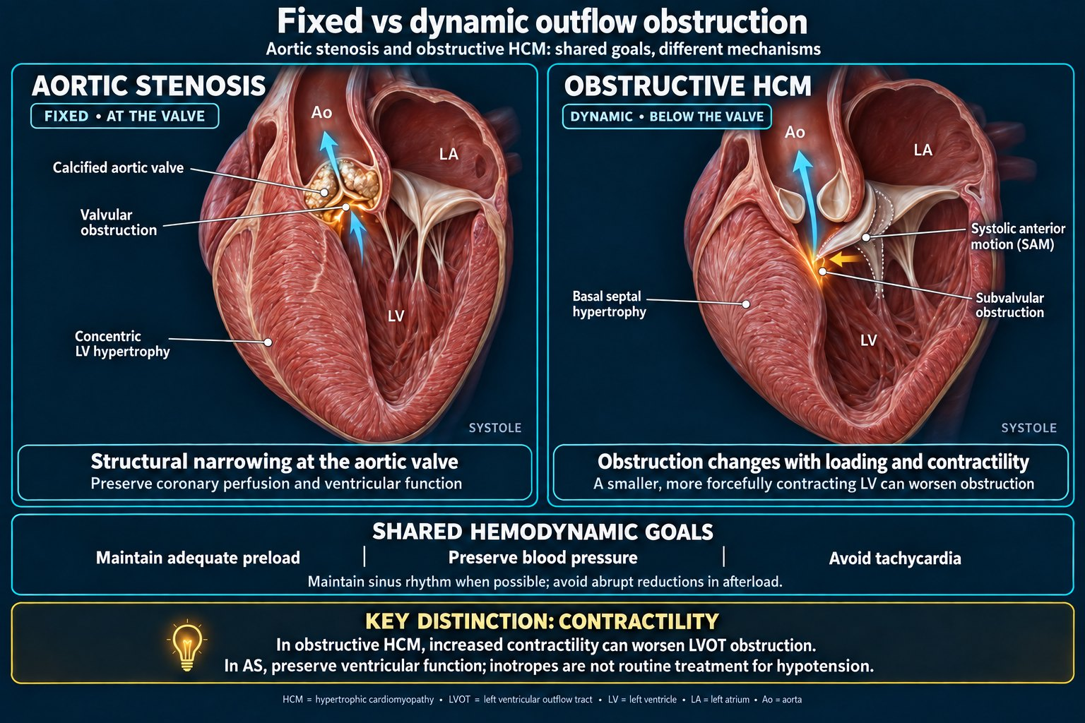
Obstructive HCM shares the goals of fixed aortic stenosis except on contractility. Hypertrophic cardiomyopathy (HCM) with dynamic left ventricular outflow tract (LVOT) obstruction shares the preload, rate, and afterload goals of fixed aortic stenosis, with afterload maintained in both, but is the reverse on contractility: increased contractility and reduced afterload worsen the dynamic gradient, so positive inotropes and vasodilators are avoided, and hypotension is treated preferentially with alpha-agonists, such as phenylephrine or vasopressin, rather than beta-agonists.1 A systolic murmur whose intensity increases with standing or Valsalva, which decrease preload, points to hypertrophic cardiomyopathy; the murmur of aortic stenosis decreases with Valsalva.20
In hypertrophic cardiomyopathy (HCM), factors that trigger dynamic outflow obstruction — positive inotropes, tachycardia, and reduced preload or afterload — are harmful and should be avoided.1 Continue beta blockers and/or nondihydropyridine calcium channel blockers without interruption, avoid hypovolemia and vasodilation, and treat hypotension by restoring volume and using alpha-agonists such as phenylephrine or vasopressin rather than beta-agonists, which worsen obstruction.1,14 Acute hypotension in obstructive HCM is a medical urgency: maximize preload and afterload while avoiding increased contractility or heart rate, and beta-blockade may be added to a vasoconstrictor.14 The distinction matters because positive inotropic agents are harmful in HCM and should be avoided to reduce the risk of hemodynamic instability.1
| Parameter | Fixed AS | Obstructive HCM |
|---|---|---|
| Preload | Maintain | Maintain (avoid hypovolemia) |
| Heart rate | Avoid tachycardia; maintain normal rate | Avoid tachycardia |
| Afterload | Maintain | Maintain — reduction worsens obstruction |
| Contractility | Maintain | Reduce/avoid inotropes — increase worsens obstruction |
| Hypotension support | Alpha-agonist | Alpha-agonist (not beta-agonist/inotrope) |
The contrast between the two lesions is specific to contractility; preload, rate, afterload, and blood-pressure goals are shared.1,2
Infective endocarditis prophylaxis
Prophylaxis is now reserved for patients at highest risk of adverse outcomes, and only before dental procedures involving manipulation of gingival tissue, the periapical region of teeth, or perforation of oral mucosa.2,12 The conditions for which prophylaxis is suggested are:2,12
- Prosthetic cardiac valves, including transcatheter-implanted prostheses and homografts;
- Prosthetic material used for valve repair (annuloplasty rings, chords, clips);
- Previous infective endocarditis;
- Specific congenital heart disease (unrepaired cyanotic lesions; repaired defects with residual shunt or regurgitation at or adjacent to prosthetic material; the first 6 months after complete repair with prosthetic material);
- Cardiac transplant recipients with valvulopathy from a structurally abnormal valve.
Prophylaxis is not recommended for nondental procedures such as TEE, esophagogastroduodenoscopy, colonoscopy, or cystoscopy in the absence of active infection.2,15 When indicated, the regimen is a single 2 g oral dose of amoxicillin 30–60 minutes before the procedure (ampicillin, or cefazolin or ceftriaxone IM or IV, if unable to take oral medication; for penicillin or ampicillin allergy, cephalexin, azithromycin or clarithromycin, or doxycycline). Clindamycin is no longer recommended for dental prophylaxis.12 Cohort data support the protective effect of prophylaxis in high-risk patients undergoing invasive dental procedures, most strikingly for extractions and oral surgery.13
Postoperative care
- Continue goal-directed hemodynamics into recovery, keeping to the lesion-specific goals, with intensive monitoring for up to 24–48 hours in AS and up to 24–72 hours in MR.2
- Watch for heart failure and pulmonary edema; longer post-anesthesia observation may allow earlier detection.1
- Treat new arrhythmia promptly. In MS, acute atrial fibrillation with a rapid ventricular response shortens the diastolic filling period and increases left atrial pressure;2 in AS, the atrial kick may contribute as much as 40% of total cardiac output.17
- Keep the heart valve team involved for patients whose valve disease meets criteria for intervention that was deferred for the surgery.1
A practical preoperative checklist
| Item | Why |
|---|---|
| Which lesion, what severity, symptomatic or not? | Determines the entire hemodynamic strategy and the risk tier |
| Echocardiogram within 12 months for suspected moderate/severe disease | Recommended before elevated-risk elective surgery to guide management |
| Does severe AS or MS meet criteria for valve intervention first? | AVR/TAVI or commissurotomy before elective elevated-risk surgery reduces risk |
| LV function, pulmonary pressures, concomitant lesions and CAD | Independent modifiers of perioperative risk |
| Invasive monitoring plan (arterial line ± PA catheter/TEE) | Continuous optimization of loading in severe lesions |
| Vasopressor chosen in advance (alpha-agonist for AS/HCM) | Hypotension must be corrected without worsening the lesion |
| Neuraxial technique modified to avoid rapid sympathectomy | Slow-titrated block with arterial monitoring in stenotic disease |
| Dynamic LVOT obstruction (HCM) excluded | Management mirrors fixed AS on preload, rate, and afterload (maintain all three) but is opposite on inotropy: avoid inotropes, which increase the dynamic LVOT gradient, and treat hypotension with alpha-agonists, such as phenylephrine or vasopressin, rather than beta-agonists1 |
| Endocarditis prophylaxis only if highest-risk and dental manipulation | Overuse is no longer recommended |
| Postoperative disposition and monitoring duration decided | Monitoring duration differs by lesion: up to 24–48 hours in AS and up to 24–72 hours in MR |
Frequently asked questions
What are the hemodynamic goals for aortic stenosis?
Maintain preload, sinus rhythm, and a normal heart rate; preserve systemic vascular resistance and coronary perfusion pressure; and avoid both tachycardia and hypotension. Treat hypotension with an alpha-agonist such as phenylephrine or norepinephrine, and manage hypertension with a short-acting arterial dilator. General anesthesia is well tolerated when rhythm and pressure are preserved.2
Should severe aortic stenosis be fixed before noncardiac surgery?
When the AS is severe and independently meets criteria for valve intervention, transcatheter or surgical AVR before elective noncardiac surgery reduces perioperative risk, and the benefit is greatest in symptomatic patients. Balloon aortic valvuloplasty may be a bridge before urgent surgery. These decisions belong to a multidisciplinary heart valve team.1,10,11
Is spinal or epidural anesthesia safe in aortic stenosis?
Neuraxial anesthesia is not absolutely contraindicated. A systematic review of more than 3,000 AS patients found no intraoperative cardiovascular collapse or mortality, with vasopressor-responsive hypotension the most common issue. The key is to avoid rapid sympathectomy — use slow-titrated epidural or carefully dosed technique with dilute local anesthetic plus opioid and invasive arterial pressure monitoring.7,8
How is aortic stenosis graded as severe?
Severe AS is defined by a peak velocity ≥4.0 m/s, a mean gradient ≥40 mmHg, or a valve area <1.0 cm² (≤0.6 cm²/m² indexed); any one criterion suggests severe disease, ideally with concordance. Low-flow, low-gradient severe AS (valve area ≤1.0 cm² with gradient <40 mmHg) is included and may require dobutamine stress echocardiography or CT calcium scoring to confirm.4,5,6,16
Which patients still need endocarditis prophylaxis before dental work?
Only the highest-risk patients: those with prosthetic valves or prosthetic repair material, previous endocarditis, certain congenital heart disease, or transplant valvulopathy — and only before dental procedures involving gingival or periapical manipulation or oral mucosal perforation. The regimen is a single 2 g oral dose of amoxicillin 30–60 minutes beforehand. Prophylaxis is not recommended for TEE, endoscopy, colonoscopy, or cystoscopy without active infection.2,12,15
How do the regurgitant lesions differ from the stenotic ones?
Regurgitant lesions (aortic and mitral regurgitation) tolerate — and benefit from — afterload reduction and avoidance of bradycardia, and are generally better tolerated than stenotic disease. Stenotic lesions (aortic and mitral stenosis) require preserved preload, sinus rhythm, and control of heart rate, and are far less forgiving of rapid vasodilation.2
References
- Thompson A, Fleischmann KE, Smilowitz NR, et al. 2024 AHA/ACC/ACS/ASNC/HRS/SCA/SCCT/SCMR/SVM Guideline for Perioperative Cardiovascular Management for Noncardiac Surgery: A Report of the American College of Cardiology/American Heart Association Joint Committee on Clinical Practice Guidelines. J Am Coll Cardiol. 2024;84(19):1869–1969. doi:10.1016/j.jacc.2024.06.013. PMID 39320289.
- Otto CM, Nishimura RA, Bonow RO, et al. 2020 ACC/AHA Guideline for the Management of Patients With Valvular Heart Disease: A Report of the American College of Cardiology/American Heart Association Joint Committee on Clinical Practice Guidelines. J Am Coll Cardiol. 2021;77(4):e25–e197. doi:10.1016/j.jacc.2020.11.018. PMID 33342586.
- Praz F, Borger MA, Lanz J, et al. 2025 ESC/EACTS Guidelines for the management of valvular heart disease. Eur Heart J. 2025;46(44):4635–4736. doi:10.1093/eurheartj/ehaf194. PMID 40878295.
- Caprio MV, De Donno F, Bisaccia G, et al. Moderate aortic stenosis: navigating the uncharted. Echocardiography. 2024;41(6):e15859. doi:10.1111/echo.15859. PMID 38853624.
- Baumgartner H, Hung J, Bermejo J, et al. Recommendations on the Echocardiographic Assessment of Aortic Valve Stenosis: A Focused Update from the European Association of Cardiovascular Imaging and the American Society of Echocardiography. J Am Soc Echocardiogr. 2017;30(4):372–392. doi:10.1016/j.echo.2017.02.009. PMID 28385280.
- Otto CM, Newby DE, Hillis GS. Calcific Aortic Stenosis: A Review. JAMA. 2024;332(23):2014–2026. doi:10.1001/jama.2024.16477. PMID 39527048.
- Tabrizi NS, Demos RA, Schumann R, Musuku SR, Shapeton AD. Neuraxial Anesthesia in Patients With Aortic Stenosis: A Systematic Review. J Cardiothorac Vasc Anesth. 2024;38(2):505–516. doi:10.1053/j.jvca.2023.09.027. PMID 37880038.
- Tai YH, Chang CC, Yeh CC, Cherng YG, Chen TL, Liao CC. Adverse outcomes after noncardiac surgery in patients with aortic stenosis. Sci Rep. 2021;11(1):19517. doi:10.1038/s41598-021-98507-6. PMID 34593867.
- Place A, Rodrigues TS, Naimo PS, et al. Peri-operative risk of non-cardiac surgery in patients with aortic stenosis: a systematic review and meta-analysis. Anaesthesia. 2026;81(4):570–579. doi:10.1111/anae.70084. PMID 41388337.
- Luis SA, Dohaei A, Chandrashekar P, et al. Impact of Aortic Valve Replacement for Severe Aortic Stenosis on Perioperative Outcomes Following Major Noncardiac Surgery. Mayo Clin Proc. 2020;95(4):727–737. doi:10.1016/j.mayocp.2019.10.038. PMID 32247346.
- Okuno T, Demirel C, Tomii D, et al. Risk and Timing of Noncardiac Surgery After Transcatheter Aortic Valve Implantation. JAMA Netw Open. 2022;5(7):e2220689. doi:10.1001/jamanetworkopen.2022.20689. PMID 35797045.
- Wilson WR, Gewitz M, Lockhart PB, et al. Prevention of Viridans Group Streptococcal Infective Endocarditis: A Scientific Statement From the American Heart Association. Circulation. 2021;143(20):e963–e978. doi:10.1161/CIR.0000000000000969. PMID 33853363.
- Thornhill MH, Gibson TB, Yoon F, et al. Antibiotic Prophylaxis Against Infective Endocarditis Before Invasive Dental Procedures. J Am Coll Cardiol. 2022;80(11):1029–1041. doi:10.1016/j.jacc.2022.06.030. PMID 35987887.
- Ommen SR, Ho CY, Asif IM, et al. 2024 AHA/ACC/AMSSM/HRS/PACES/SCMR Guideline for the Management of Hypertrophic Cardiomyopathy: A Report of the American Heart Association/American College of Cardiology Joint Committee on Clinical Practice Guidelines. Circulation. 2024;149(23):e1239–e1311. doi:10.1161/CIR.0000000000001250. PMID 38718139.
- Nohria R, Romaine A, Garcia-Sampson G. Infective Endocarditis: Diagnosis and Treatment. Am Fam Physician. 2026;113(2):145–152. PMID 41839106.
- Coisne A, Lancellotti P, Habib G, et al. ACC/AHA and ESC/EACTS Guidelines for the Management of Valvular Heart Diseases: JACC Guideline Comparison. J Am Coll Cardiol. 2023;82(8):721–734. doi:10.1016/j.jacc.2023.05.061. PMID 37587584.
- Sarwar MF, Rodriguez-Diaz C, Dabski M, et al. Anesthesia for cardiac surgical procedures. In: Gropper MA, Eriksson LI, Fleisher LA, et al., eds. Miller’s Anesthesia. 10th ed. Elsevier; 2024:1643–1644. Source of the diastolic dysfunction and rise in LV end-diastolic pressure that follow concentric hypertrophy in aortic stenosis, and of the atrial kick contributing as much as 40% of total cardiac output.
- Khanna S, Hargrave JM, Abraham A, et al. Anesthesia for cardiac surgery. In: Cullen BF, Stock MC, Ortega R, et al., eds. Barash, Cullen, and Stoelting’s Clinical Anesthesia. 9th ed. Wolters Kluwer; 2024: chapter 39, Table 39-8. Source of the rhythm goal in aortic stenosis: sinus, with cardioversion or rate control considered in a nonsinus rhythm.
- Pitkänen M. Spinal (subarachnoid) blockade. In: Cousins MJ, Carr DB, Horlocker TT, Bridenbaugh PO, eds. Cousins & Bridenbaugh’s Neural Blockade in Clinical Anesthesia and Pain Medicine. 4th ed. Lippincott Williams & Wilkins; 2009:214. Source of the concern that in marked aortic stenosis the sympathetic block of spinal anesthesia can suddenly lower systemic vascular resistance and coronary perfusion, and that a limited block or a carefully titrated continuous technique can be used.
- Wijeysundera DN, Finlayson E. Preoperative evaluation. In: Gropper MA, Eriksson LI, Fleisher LA, et al., eds. Miller’s Anesthesia. 10th ed. Elsevier; 2024:830. Source of the murmur maneuvers: standing and the Valsalva maneuver decrease preload and increase the murmur of hypertrophic cardiomyopathy, and the Valsalva maneuver decreases the murmur of aortic stenosis (Table 28.9).
Disclaimer. This page is an educational reference for licensed clinicians. It is not medical advice, does not establish a clinician–patient relationship, and does not substitute for individualized assessment. Anesthetic management must be tailored to the specific patient, valve lesion, severity, symptom status, procedure, and institutional resources. Verify all drug doses and thresholds against current guidelines and package inserts.
This page was written and cited by the CRNA who builds Helix Anesthesia, a point-of-care reference built for the operating room — surgical cases, drug dosing, regional blocks with labeled ultrasound anatomy, and crisis checklists, all cited and available offline.
The guidance above is cited to the primary literature listed rather than to the app’s own case text. The sourcing standard is the same either way, and it is documented at how we source clinical content.
Built for the anesthesia machine, not the desk. It opens in one tap, works with no signal in an OR that has none, and shows the citation on screen beside the number rather than making you go and find it.
Free tier, no card: every calculator, every crisis checklist, the full drug library and glossary, three complete regional blocks, five complete surgical cases, and 15 AI questions a month.
Get Helix Anesthesia How we source itSign up for our newsletter and special offers. New answer pages, improvements to the ones already here, and the occasional offer. Nothing else, and not often.
Hemodynamics
- Spinal anesthesia in the elderly — start here
- Ondansetron and spinal hypotension
- Vasopressor equivalents
- CABG anesthesia
- Surgery timing after a stent
- Etomidate and long-term outcomes
- Pulmonary hypertension
- Intraoperative hypotension
- IV agents: hypertensive emergency and AF
← Clinical references for anesthesia providers — the full index for clinicians. · ← What to tell your anesthesia provider before surgery — the plain-language index of these guides. · How we source clinical content · All clinical answers